
Speaking at the Return to Play Conference 2014 in Melbourne, Dr Jowett revealed that ACL injuries do not only affect professional sports stars, they also impact the community level athlete.
This leads to broader impacts on employment and the ability to work in a physical environment and go well beyond the cost of surgery and rehabilitation.
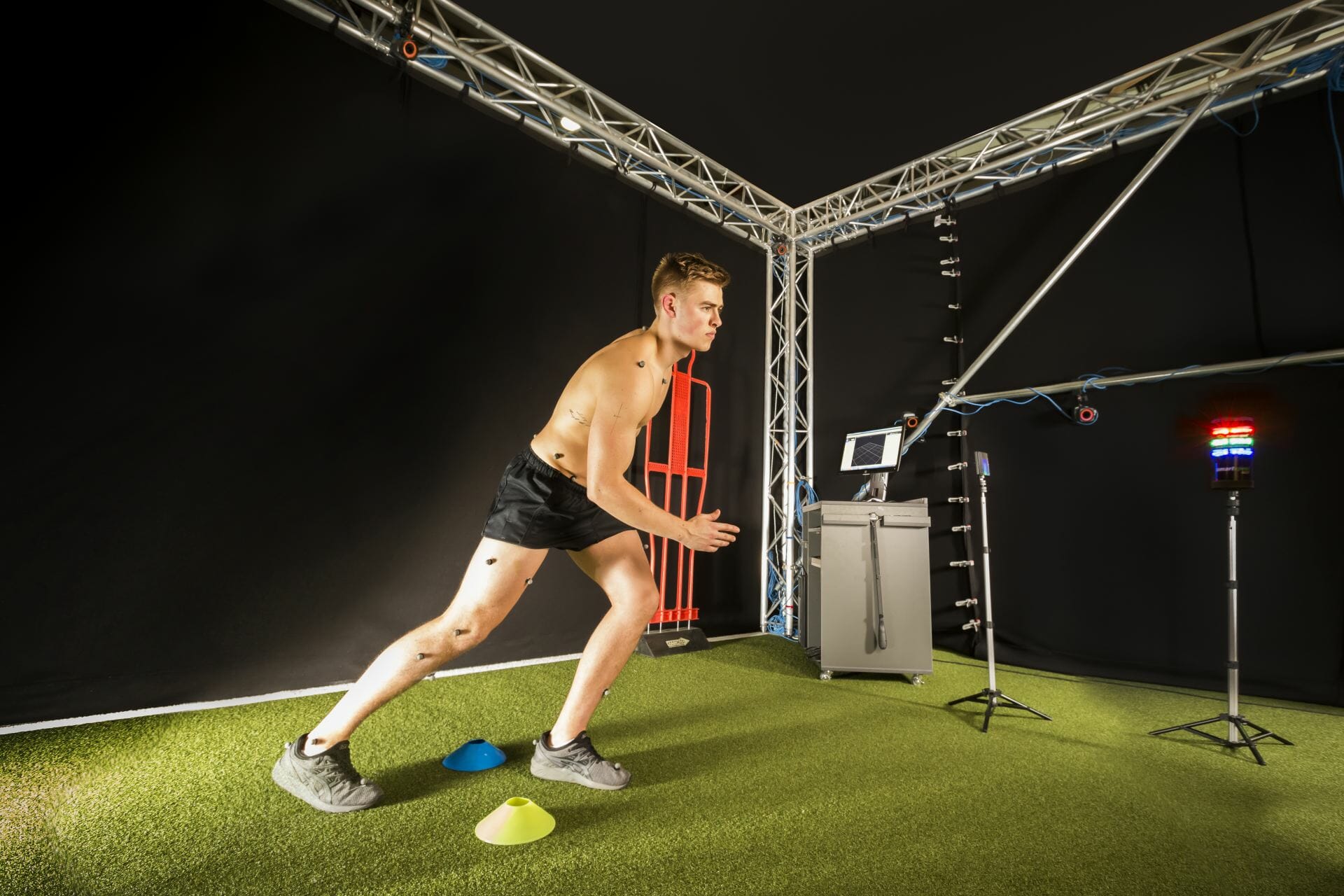
To help study the prevention of ACL injuries and rehabilitation, there is a requirement to understand how often they occur, and not just at the elite level either where there is a lot of data available.
For example, ACL injuries are 4-6 times more common for Australian international women footballers compared to their male counterparts, stats that correlate with data at an international level however according to Dr Jowett there is no data as to what happens on the levels below.
The gaps exist where people do not get ACL injuries treated, or they are simply not recorded.
In general, there appears to be a change in the modern lifestyle, there is less unstructured play. Kids nowadays don’t really climb, they play and train to structured sports programmes.
Physical Education in the school system should be more about unstructured movement rather than competitive sports.
A change in direction is needed at an administration and programme level. A register of injuries at all levels would help the collection of data while a prevention programme at a younger level would help stem the number of ACL injuries.


 Patellar tendinopathy is a condition we see commonly in clinic and can cause athletes from many sports a lot of discomfort and frustration. The Patellar tendon runs from the patella (kneecap) to the tibia (shin) and transmits the force from the quadriceps muscle to the lower leg to allow you to straighten your leg against resistance.