Back Pain Rehabilitation

‘Low Back Pain: Facts, Fallacies & Self-Management’ with Katie Gill Senior Lumbar Physiotherapist

Watch this video of Katie Gill, Senior MSK Physiotherapist, at UPMC Sports Surgery Clinic in Santry, presenting on ‘Low Back Pain: facts, fallacies & self-management.’

This video was recorded as part of SSC’s Online Public Information Meeting, focusing on Back pain & the Spine.
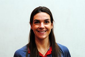
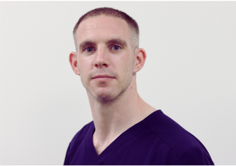
Katie Gill

Firstly, a bit about my background I qualified as a physio from UCD in 2011. I worked in private practice for several years in Dublin before moving to London to undertake my Master’s in advanced practice MSK.

I worked in the NHS for a couple of years in a mixture of acute persistent or acute and chronic services, and I moved back to Santry in 2018 to start my current role with UPMC Sports Surgery Clinic.

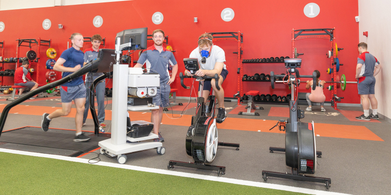
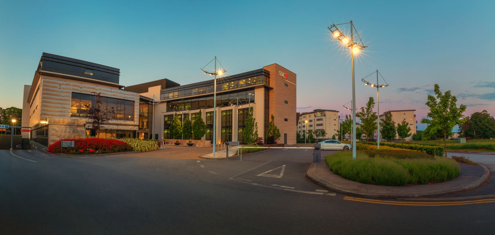
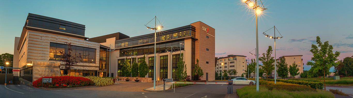
You can see in the top picture that this is the main hospital where all the orthopaedic surgeons work and where all the operations occur. The picture below is Sports Medicine, where I work; it’s located around the corner but still part of the hospital. Our Sports Medicine Team covers a good mix of services in this building. We have Physiotherapists, Strength and Conditioning Coaches, Nursing Administration and Sports Medicine Consultants.

I am one of eight Physiotherapists and S&C Coaches doing research alongside clinical practice. By doing this, we can better our patients quicker, contribute to the overall medical body of knowledge and ensure excellence in our clinical care.

The aim of the presentation this evening is to discuss Back Pain. First, we will look at the background of low back pain and then at treatment guidelines, management, and advice from a Physio’s perspective. In the next presentation, Dr Matthew Cosgrave will give his views on the same from a physician’s perspective.

We’re going to look at some myth-busting and look at some sign postings for some resources, and there’ll be time for questions at the end.

Just as a note that the presentation shouldn’t be used for individual advice for your specific queries or back pain, and we’re kind of generally talking about low back pain this evening, so if you have any specific questions or concerns and I’d advise you to seek the advice of your physio or healthcare professional.

I’m sure some of you who are listening might be frequent flyers into physio and know what it’s all about; others might be listening to this thinking, I have no idea what to expect when I have back pain and attend a physiotherapist.

Firstly, your Physiotherapist will ask you to provide them with a history of your back pain. When did it start, how long has it been ongoing, and are there movements that make it worse or better?

Then a physical exam will examine how your back and hips move, and some strength measurements will be taken.

At the end of the session, there will be a conversation where you can ask questions, and we will go through some advice on treatments.

So physiotherapy sessions are usually divided into three sections, the history, the exam, and the treatment.

As healthcare professionals, physios are obliged to work within evidence-based treatment guidelines. This means that we have to offer and be up to date with all the evidence and research coming in through our field. If you’re going to see a physio, you should ensure that they are CORU registered to ensure they are keeping up with evidence and standards.

So the first thing is back pain can be really scary, but it doesn’t need to be. I hope this presentation will help alleviate some of the fears associated with low back pain.

I find with patients that their fear often comes from not understanding the source of their pain.

If you find yourself in this situation, I recommend you tell your Physiotherapist what is worrying you at the beginning of your consultation.

Now we will keep returning to this notion of the fingerprint. While back pain can be very straightforward to treat, it can also be complex. Think about your back pain as you would think of your fingerprint.

It is very specific to you as an individual. We will develop this idea later in the presentation but remember that your treatment needs to be tailored to your specific needs.

Looking at the background of low back pain, the picture at the top is from a paper published in the Lancer, a very well-respected Journal. In 2018, they did a vast series of documents highlighting how common and prevalent low back pain is.

It is the leading cause of disability globally, and at any point in time, about 550 million people are experiencing it at any one time, which is vast. Most people will experience it during their lifetime at some point.

I hope talks like this will help put the correct information out there and that people learn that low back pain isn’t necessarily something to be frightened of.  It is unpleasant, but it is very treatable, and people should know what to do and not do when they suffer back pain.

The biggest thing to take from this slide is that if you’re playing a numbers game, most cases of back pain must settle within six weeks. Somewhere between 80% and 90% of episodes will settle within six weeks if you do nothing without any treatment from your GP or Physio. That’s an empowering statistic because your back is always working behind the scenes to settle that pain.

Another thing to remember is that serious causes of back pain are extremely rare. Less than 1% of patients have something that is serious or requires ongoing management or investigations or has a serious underlying cause. So it is very rare.

But why do so many people feel there’s something seriously wrong?

When I talk to my patients, they believe something is seriously wrong with their backs, as their pain is excruciating. Back pain can be really sore. In the best cases, you are uncomfortable, unable to do the things you want to do. In worst-case scenarios, people experience excruciating pain so naturally, believe something to be seriously wrong.

I would like to reassure you that the pain and damage aren’t the same and that you can be sore but safe.

We break back pain into two categories. Specific and non-specific.

Specific back pain relates to 5% to 10%  of cases depending on which research you read. Specific means you can point to something or know the cause of your pain. It might be an infection because say you’ve had surgery. If there’s an infection in the back that’s a specific cause, or you’ve had a fracture in your back that’s a specific cause, we know exactly what’s causing it.

Everything else is called non-specific. So 80% to 90% of people fall into this category, and it’s unsatisfactory. It’s extensive, and I will explain why it’s so broad, but most people are in that non-specific low back pain category.

Timeline is another way to categorise back pain. Acute backpain refers to the first six weeks and persistent back pain for six weeks and onwards.

Why are so many people in that non-specific low back pain category and why can’t we tell exactly what’s causing their back pain?

Part of the reason is due to the anatomy of the lower back.

if you look at my finger here you can localise my anatomy around my finger, But if you look at the picture on the left the yellow bits are the spinal nerves and in between are joints. Two joints might be a centimetre apart on the left and a centimetre apart on the right and there’s a lot of anatomy within the same area. All of these body parts have a nerve supply so your skin, your muscles, your ligaments even the nerves of your discs have a nerve supply and they’re all very close together.

So if you’re lifting, carrying, moving or bending forward you’re going to be moving all of them so it’s very hard to say well look it’s absolutely one or the other causing the pain.

If you look at the picture on the right in terms of your lower back you will see the anatomy is very complex. There are a lot of muscles covering this area so we used to think we were able to diagnose precisely the cause of it every time – it’s just that the anatomy is too complicated to let us do that.

Discs have a nerve supply, so some problems can be very painful, and some discs can have degenerative changes. It’s just like getting arthritis in our knees, arthritis of our hips or growing hair and wrinkles. It’s part of our ageing process and doesn’t necessarily cause any pain.

Think about how your body adapts to that. If your discs change slowly over time, your body doesn’t see that as a problem. But if somebody has a sudden injury,  they fell down the stairs, and there was too much pressure or force, causing a sudden change, that disc can be painful.

Ten years ago, a significant study was conducted on 3000 people on this subject. A cohort of people between the ages of 20 to 80 who were not suffering from back pain was given an MRI.

MRI showed that 30 % of people in their 20s had disc bulges but no back pain. The figure was 50% for those aged 50 and over which is a significant amount of disc bulge with no symptoms of back pain.

So the MRI findings need to fit in with your own personal history and with your clinical patterns. You are subjective, and I think getting advice from your Physio or Healthcare practitioner around that is essential in order to interpret them correctly.

I spend a lot of time with people who are just given reports and understandably have a long list of things that come up from an MRI, and a lot of it is like grey hair and wrinkles; it’s part of how the body ages and not something to be worried about.

I like to focus on evidence-based exercises and treatments for low back pain. You may have heard of NICE or The  National Institute for Health and Care Excellence.

They are a huge research body that provides guidelines for rehabilitating things like low back pain or obesity, and smoking.  The same applies to Cochrane.

The following guidelines have been approved by these two bodies.

They are divided into Acute Low Back Pain and Persistent Low Back Pain.  As a reminder, Acute Low Back Pain is what we have from zero to six weeks, and this table is taken from the paper I referenced earlier on, and I have the reference for you at the very end of this presentation.

What is the evidence advising during the first six weeks?

The first advice is to remain active, but look at and pacing yourself, and manage what you need to do.  For example, if you have an irritated back in the first six weeks, this is not the time to try and run a personal best or do a big clear out in the house or swing the grandchildren around. It’s about doing what you can, pacing yourself, and maybe keeping yourself moving without overdoing it.

The interesting part here is exercise therapy has limited use in selected patients, so from a physio point of view, I tell people to do the movement things that they enjoy. I try to give them some basic exercises if needed. It’s more like telling people they’re sore but still safe, and I try to get them back into things they need to do.

You’ll know they’re at the very bottom for things like manual therapy, spinal manipulation massage, and Hands-On work should be in conjunction with the education and remaining active, so it shouldn’t be done by itself it’s if you’re seeing a Physio and they’re doing some Hands-On that’s fine. However, it still needs to come with advice and education as well.

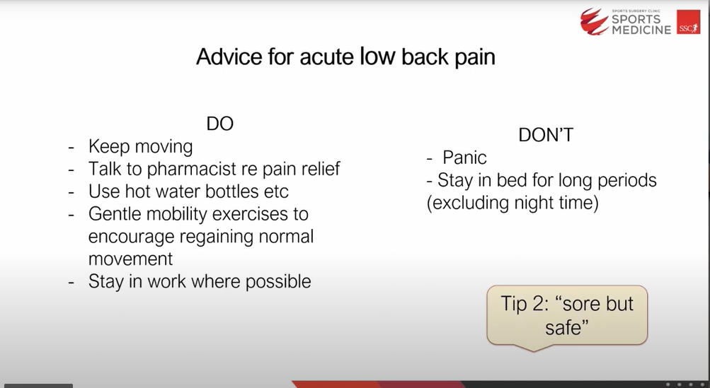
To recap, here are the dos and don’ts for low back pain. If you want to take a picture of this slide, I don’t mind.
Low Back Pain SSC Santry

So do keep moving. You could talk to your pharmacist about pain relief; you can use hot water bottles and gentle mobility exercises to encourage normal movement. It is really important to stay at work where possible and don’t panic. Back Pain can be very sore but we want to try to get the body out of this fight or flight mode and back into a more relaxed state.

The advice 20-30 years ago was to stay in bed. Try not to stay in bed. Get up and move around every 20 minutes to half an hour. If you’re sitting, resting, standing up, sitting down, walking to the kitchen and back, and just trying to keep yourself moving.

I would say to patients at this time that they are sore, but they are safe.

Can pain be a good thing?

I’m sure everyone thinks that there’s no way pain can be a good thing and that I am actually like Pinocchio and that I’m telling lies, but I promise you I’m not especially in the acute phase of back pain.

Imagine your pain is your body trying to talk to you asking you to change your behaviour. There are a group of people with this genetic mutation who cannot feel pain. This sounds wonderful but their bodies don’t have a way to tell them if they have an infection or have broken a bone.

Their mortality rate is relatively high, so we need our pain system to work, and tell us what we need to pay attention to. It’s you’re body letting you know that we’re not going to go to the gym today and that we will do more gentle exercise. So our pain system must be there because it keeps that warning system in place, which is good.

Persistent Back Pain.

Persistent low back pain is when the pain is there longer than six to twelve weeks. If you are suffering for this amount of time I would recommend you visit your GP or Physiotherapist because you’ve suffered for long enough and there are lots of treatments available so there is no need to suffer.

The advice or evidence here is slightly different, so the advice is still to remain active and educated is still essential.  However, exercise therapy now becomes the first treatment. It’s as crucial that its used in conjunction with manual therapy, and actual hands-on treatment which is secondary. Exercise advice and education are all the first things that need to be part of your physio program, and some hands-on work can be used, but it needs to be done in conjunction with the above.

What are the challenges in treating persistent low back pain?

We are going to remember that this is like your fingerprint and pain has a context so the best example I can give for that is imagine that you’re having a really good day that you fall up the stairs at work and it’s really funny you think oh my gosh look I’ve landed on my knee and I’ve hurt myself. Imagine you’re late for work and you’ve got soaked going in from the car you fall off the chair land on your knee and everything is the worst you’re really sore.

The same thing has happened but the context is different so our thoughts, our feelings and our beliefs can shape the way we shape our pain experience remember that if we have an irritated or grumpy back that back is attached to a person in their lived experience so and all of our lived experiences are completely different. I will expand on this in a minute.

The paper I referenced earlier shows different factors can be at play in terms of feeding into low back pain. Things like genetic factors and biophysical factors. So there is strong evidence that people with low back pain have some changes in their strength and coordination. There are other comorbidities like anxiety, depression, social factors and things like stress that can add to the causes of low back pain while not necessarily being the root cause.

Remember that our pain and why low back pain has been persistent and more complex to treat is because everybody is going to have a different interplay or some people might have absolutely no stresses in life and none of that going on, and really all we need to focus on is the muscles and strengthening. For other people, the strengthening is not a big factor we need to focus on pacing and management so it really just depends.

Why is exercise important for treating back pain, and why is it recommended?

The American College of Sports Medicine has published guidelines on physical activity for adults. This is what we all should be doing anyway as healthy adults. Ideally, everybody needs 150 minutes of moderate-intensity exercise or 75 minutes of vigorous-intensity a week. In addition, we should be building our strength on two days or episodes a week.
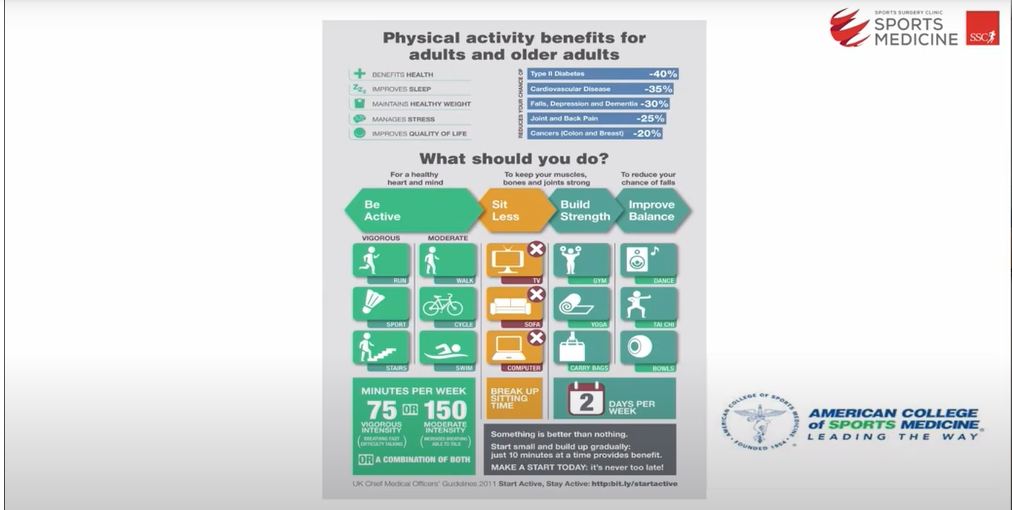
American College of Sports medicine Physical Activity Guidelines

This is quite a high amount of exercise, but it’s what we should be doing not just to be fit but to maintain a baseline level of health. So sometimes, patients with low back pain can miss out on the benefits of general exercise because they’re sore.

Why do we recommend our patients undergo strength or resistance training at SSC?

We encourage our patients to do some strength or resistance training here in Sports Medicine for several reasons.  In this slide, the bottom left is a picture of a cross-section of the human back.

The two dark circles at the very bottom are the back muscles. Resistance training can improve the amount of fat within a muscle. Another reason for promoting resistance training is it is very good for maintaining joint range of motion in our spine, inner hips and all over the body. It is excellent for building bone strength as well as building muscle strength.

Our lives require us to have a level of strength so if you think of pushing a shopping trolley, opening a heavy door, lifting grandchildren or walking to work, all of those things require us to have a certain level of strength, so it’s being able to meet the demands of your day-to-day tasks as well as the positive impact it has on our mental health.

Our Physiotherapy Department at UPMC Sports Surgery Clinic takes self-referrals. You can phone us on 01 5262040 or email [email protected] for further inf

Do you have any recommendations for exercise that a person can do to help relieve pain from spinal arthritis in all three parts of the spine?

yeah I think so there’s some um there’s lots of great questions coming in and we’ll try to answer as many as we can but certainly with marks in terms of exercises like we spoke about in in the Talk looking at things that you enjoy so try to sometimes pay people with spinal arthritis they find that extremes of movement so it might be do yoga but maybe avoid an extreme back bend or stream front Bend if that doesn’t feel right to you so what I’d say in terms of guidelines and pain from spinal arthritis is that the odds nip or pinch with pain is okay if an exercise feels like you know you really don’t feel good afterwards it’s really making that pain worse that one isn’t for you so examples of things with spine arthritis there’s no limits you could run you can do yoga you can swim really looking at what trying to meet those recommended guidelines that I spoke about earlier try to get your enjoyment in but there’s no Bible or gold standard of the thing you absolutely have to have to do and that links in with Maureen’s question which that she spoke about having um some fusion due to arthritis so what that will mean if somebody has lots of arthritis down at the bottom of their spine some of the movement around that region isn’t going to be as good which we’d expect but what if you what you want to try to do is maximize the movement that you do have and maximize the areas above where the fusion is or where the arthritis is and below so it might be making sure you’re getting good hip range of motion and making sure you get good Lumbar and thoracic range of motion so from a treatment an excise point of view Mobility work, yoga will be nice and resistance training would be nice, medical questions I’ll leave I’ll leave for Matt, for that but it really is like our guidelines from earlier on is trying to meet recommended guidelines, do something that you enjoy and if something doesn’t feel right or is painful or if you want more specific advice I think I would chat to your own physio or GP.

A patient has been diagnosed with scoliosis-like species in the past year with grade one slippage of the vertebrae onto the L5 S1 nerve, causing severe back pain and groin weakness. He has been going to Physio and a chiropractor with limited results.

okay so I think my guess is follow-up questions to this patient, and we’re trying not to answer this more broadly because I haven’t assessed them myself. Still, certainly, it depends if you’ve been giving us a good go with a physio or a chiropractor treatment. Indeed, if it’s been going on for a while, I think Hands-On work from a physio or a chiropractor probably sounds like it’s not working, so I think a different approach might be worth there going back to the GP and looking for a second opinion.  So what all those terms mean for anybody who’s not familiar with them is just that there’s a little bit of movement of one of the vertebrae, which is putting pressure on a nerve. Some injection options and others depend on how severely you’re impacted from a particular exercise point of view, so certainly, I think if it’s not working with a physio or chiropractor, my first question is, what type of things are they doing with you? If it is more Hands-On, I would put a pin in that go back to the GP looking for an opinion and move from there.

I was wondering how long it takes the muscle condition to deteriorate within an activity, especially in the lumber area, okay?

That’s a great question, so I wonder whether that came from seeing the cross-section of the spine in the presentation earlier. I showed pictures of where there were lots of fatty infiltrates that can level of infiltrate for years, so that’s not something that would happen over weeks or a month, so that level of infiltration when someone is very sedentary takes years, so in terms of deconditioning so you can lose some muscle strength within two to three weeks. Still, you can get it back again really quickly, so I don’t want people to worry or necessarily that if they’ve been on holiday or if you’ve been, you know, unwell for a period that you know your muscle strength completely goes it doesn’t and what you want to try to do is to get your muscles in good condition as best you can. So you’ve noticed the difference in, say, if you were in the gym and what you would lift if that’s a good level of muscle strength. It might be well you know I lifted my grandchild before I went to my six week holiday and have come back to I feel they are bit heavier you might notice some impact in your function after several weeks a very significant deterioration where there’s a tremendous amount of fatty infiltrates that takes years. Still, you can notice lower grade changes within a couple of months. I hope that answers that.

For further information on back pain or to make an appointment with one of our Physiotherapists, please contact [email protected]
Book Now 1

‘Sports Medicine and Low Back Pain.’ -Dr Matthew Cosgrave

Watch this video of Dr Matthew Cosgrave, Consultant Sports & Exercise Medicine Physician. Presenting on ‘Sports Medicine and Low Back Pain.’

This video was recorded as part of SSC’s Online Public Information Meeting, focusing on Back pain & the Spine.

Dr Matthew Cosgrave SSC WEB.png

MB BCh BAO MFSEM (IRL & UK) MSc (SEM)

Dr Matthew Cosgrave is a Consultant in Sports and Exercise Medicine. A graduate of Medicine from Queen’s University Belfast in 2011, Dr Cosgrave undertook an MSc in Sports and Exercise Medicine at The University of Bath before completing specialty training in General Practice in Belfast in 2018. Dr Cosgrave worked as a general practitioner with a specialist interest in musculoskeletal and sports team care and as a specialty doctor in pre-hospital medicine before moving to Dublin to complete Higher Specialty Training in Sports and Exercise Medicine in Ireland.

Hello everybody, good evening; I hope you’re enjoying the evening. I’m delighted to be able to join you and discuss the sports medicine doctor’s role in managing low back pain. My name is Dr Matt Cosgrave or Matthew, and by introducing a little bit of my background, I studied in Belfast at Queens University. I originally trained as a GP, working through various Specialties in the NHS. Then I moved down to Dublin to pursue a career in sport and exercise medicine and did my speciality training in Dublin outside of working in the clinic here at Santry. I work with a number of sports teams, but over the last three or four years, I’ve had the pleasure of mainly working with the Irish Women’s National Rugby team and, over the last couple of years, with Connacht rugby men’s senior team. From the point of view, what I hope for you to get out of my talk today is to answer these three questions do I need to see a sports medicine physician? Do I need an MRI, and do I need an injection? Before we get into things, I’d like to reassure you all that when we talk to patients, these are the three most common emotions that they present with in relation to low back pain worry over some underlying disease process or pathology that maybe they’re missing, confusion over often why their pain isn’t getting better or why their pain comes and goes unpredictably or apathy which is really a down to a lack of motivation around managing their pain and that no matter what they seem to do they’re always suffering. Interestingly enough, these three CM emotions are often what positions physiotherapists or General Practitioners experience with low back pain because it can be tricky. Still, hopefully, I will try and uncover some of the Hidden Truths Behind that today.

I don’t think we help ourselves by calling ourselves Sports Medicine Physicians; this is a common misconception that the only people that we treat are people who do sports or athletes; we are musculoskeletal Physicians, so yes, we do look out for teams, yes we do look after athletes, but we look after any active individual which from the point of view of day to day that’s the general public and the vast majority of the patients that we see in care are general public and are not high-flying athletes in any shape or form. About eighty percent of people at some stage in their life will experience low back pain. Interestingly, about eighty percent of these cases will improve on their own or with very little treatment within about six weeks. My rule comes into it whenever we have this huge chunk of 20 percent who are not improving over the three to six weeks or the three-month to six-month period, which can be a substantial number when you cross when you break it all down.

Really what the sports medicine physician does is considers do we need to order other investigations now this may be a scan of some description a CT an MRI or it may be some blood tests really the main role for us is to confirm a diagnosis often once we have the diagnosis actually managing the problem can be quite straightforward sometimes when we’re in the dark about the diagnosis we try and we almost have a trial and error process but if we can confirm a diagnosis it’s often fairly straightforward to get on a plan to recovery excluding severe pathology is a big part of this and this falls into the worry side of things for patients a big concern that patients often have is there some sort of underlying disease process such as cancer and that is extremely rare when it comes to low back pain but nonetheless when back pain has been going on for a long time it’s always something that we have in the back of our mind and it’s always something that we make sure we exclude we’ll also review the treatment plan that the physiotherapist or the GP has already put in place and we’ll see can we add to that we rarely change things significantly but we’ll make small adjustments and then finally we think about treatment adjuncts so rehab is a huge part of what we do here in the clinic but there’s always options for some additional help and often for us that falls in the way of injection therapy which I’ll discuss for you in a little bit further detail later.

The simple answer is no, not everybody needs an MRI, and most patients with low back pain don’t require an MRI. This great graphic explains why we don’t all need MRI. These are findings on individuals who are asymptomatic so people with no back pain have a variety of changes, and the percentage of patients who have changes increases with age; just for one example, this degeneration in this top area which is often reported on scans and often causes concern for patients nearly 100 percent of patients in their 80’s will have this degeneration but even more interesting at the age of 30 one in every two patients will have some degree of disc degeneration on their scan even if they are asymptomatic. Hence, a scan is not always necessary.

So when is the scan necessary?

We use scans; this is what the sports medicine position is looking for if we go clockwise from the top left; this is a condition called sacroiliitis. This is an inflammatory process where you get some inflammation in the base of the lumbar spine where the sacrum, a little triangular bone, fits into the pelvis; we look for sacroiliitis because it doesn’t normally respond to Conventional treatment. Usually, it’s caused by some inflammatory process in the Blood, and therefore we often need to give medication to reduce that inflammation in the blood, which will then reduce the inflammation in the joint; this is not very common and but when it does present it tends to be in the younger person. If we move over to the right, then we have this condition called facet irritation; this is a lumber spine from the side; the square blocks on the left are the vertical bodies, and they sit on top of intervertebral discs the vertebrae then communicate with each other and the back through something called a Facet Joint now this Facet Joint like any joint in the body a knee, a hip they can become irritated and inflamed so when we look at MRI scans of backs or patients who are not improving we’re often looking to see are there any individual joints that are irritated or inflamed that could be contributing the symptoms and therefore is there a Target for some injection therapy. In the bottom left, this is the view of a spine in cross-section, so this is like I’ve taken you lying down and then chopped you along, and I’m looking down the body; this pointy bit at the back is called a spinous process, and this is the area that we feel when we feel down in the back. In patients, again usually young patients who are very active and who do sports that involve a lot of bending off the back, so gymnastics or tennis, there is a risk of developing something called a porous fracture or a stress fracture, so in the younger patient especially this is always something that we want to exclude. Finally, which you’re probably most familiar with when it comes to low back pain is this condition in the bottom right called disc degeneration; the black circle in the center is the spinal cord the yellow marks to either side are the nerve Roots. When you get a disc bulge or a disc protrusion, the center of the disc, a jelly-like material, pushes out a little bit, and it causes the disc to bulge the disc itself doesn’t move. It creates a little bit of a bulging on the outside edge; now, this bulge can do a few things; one is it can create irritation around the nerve. The other is if it’s bad enough, it can put some pressure on the nerve, and then if we think about treating this often, what we think about doing is delivering some injection therapy, usually a steroid in and around this area to reduce inflammation and irritation and to reduce any pressure that’s on the nerve itself. Hence, these are the four main reasons why we do MRI scans, but we know specifically what we’re looking for; we’re not doing the scan and hoping that it gives us the answer we’re doing the scan because we think that we already know the answer and we’re trying to confirm or exclude that. From a disk point of view, I just wanted to give you a little bit more detail so from a disc bulge point of view, the reason this is important this is a spine on the left from the side this is the spine on the right from the front these grey areas that are running down the side and then are coming out on either side on the image on the right these are nerve roots your nerve Roots Supply your sensation and your part to different levels along your lower legs so when you come into us with symptoms what we do is we try to correlate whereabouts on the leg front, or back your symptoms are and then when we do your MRI scan we try to look at the disc at that level to see if it’s causing some pressure around the nerve root.

In many cases you will have disc bulging it doesn’t relate to your symptoms so you may have disc bulging at this L1 L2 level but your symptoms may be coming from L5 or L4 for that reason MRI’s are not useful in that they can create confusion for patients and sometimes for Physicians as well because we see disc bulging but it doesn’t marry up with what’s going on in the history .finally sorry I want to make a quick note on something which Katie has probably already discussed which is a degree of fatty  infiltration in the low back when we are less active we see a change from the left hand side where the muscles are nice and grey kind of like a fillet steak to on the far right hand side when the muscles are not being used because other areas of the back are having to work harder for example the facet joints then the back becomes weaker and we see this fatty infiltration if we see this on a scan it’s a very good indicator to us that you’ll respond well to rehab so in summary from an MRI point of view we’re looking to exclude significant disease we’re trying to correlate what we see and what you tell us with what we find on the image or on the MRI scan it’s really not guesswork and we’re not asking for the MRI to give us the answer.

Finally, we’re looking essentially if there is an area for injection. Hence, if we see a disc bulge or a Facet Joint, that’s irritated. If it fits with where you’re sure and where we examine you, then that’s usually a good Target for us to inject the injection is only part of the treatment plan, which I’ll discuss and that brings us on nicely. Do I need the injection? Injections are not always straightforward, and often patients are a bit apprehensive about them.  injection treatments are put into two major areas; one is for nerve root impingement, so if we have this disc pathology or there are other causes of nerve management, but if we have nerve root impingement, we’ve got two options, we’ve got a coral epidural injection, or we’ve got a more sophisticated CT guided nerve root injection again we’re correlating our MRI findings based on where your symptoms are. We are deciding on what level you need the injection.

Sometimes it’s not always clear exactly which level sometimes you might have a disc bulge at a couple of levels and sometimes you might have symptoms that correlate with a couple of levels when that’s the case we’ll often do a call to epidural injection this injection is done right down at the base of the spine we inject some steroid and it flows up along and around the nerve roots and it beads the nerve Roots at those lower levels in steroid and it reduces any inflammatory or Earth and change around there it’s a little bit of a broad brush stroke and we’re hoping to tackle a number of different areas in one go the CT guided nerve right injection however is much more selective we do that when we are very confident that the image that we see on MRI fits perfectly with the way the patient comes in now this is not always the case but when we have this sort of scenario you get much more relief from us a selective nerve root injection than by doing the broad brush stroke of what we describe as I called epidural injection technically we call the epidural is slightly easier than the CT guided nerve root we perform the seat they call epidural injections in the clinic here as Sports Medicine Physicians we do this under ultrasound scan the selective nerve roots however we have to refer to our Radiology colleagues in the main hospital for this because it’s slightly more sophisticated and we want to be exactly precise about where were placing that steroid.

The next group of injections then is for the joints I mentioned to you earlier about this condition, sacroiliitis or inflammation around your sacrum again, normally the way that we treat this is with some medication to reduce inflammation in the Bloods, but on occasion, if you have it on one side it can be because the joint is irritated without inflammation in the blood, in that case, a steroid injection over that area can be very helpful again we do this in the clinic here. We use an ultrasound scanner to guide where we place the needle.

Finally, then is the facet joint injection, the Joint between the different levels of vertebrae same principle as a knee joint, like a hip joint. If it gets inflamed, they often respond very well to cortisone steroids; it is not the main way of treatment; it’s an adjunct, so we try to improve your symptoms a little so that you can then progress on and do whatever rehab is necessary.

Are injections necessary?

Not always, but we use them when patients are not improving or deteriorating in some shape or form. We only do them if the symptoms that you’re telling us or the findings that we have on examination match up with what we see on the MRI scan; if they don’t match, there’s something else happening, and that’s rarely the case. Finally, we do them if your symptoms stop you from doing good rehab. Suppose you’re too sore to rehab, and we have a focus on injection. In that case, these can be extremely helpful in trying to get you some pain relief so that you can do the strength work it helps to take the pressure off the back it helps to get you moving more naturally, get you feeling more comfortable and then get you back to living a healthy and happy life.

Very straightforward self-referral, GP referral or physio referral, and you can forward an email or a referral to [email protected], or you can call us at this number 015262030. You can have a discussion with one of the staff at the desk, and they’ll give you more direction about how to get in contact with us. I hope you found that helpful there’s a lot of information in there. You don’t need to take it all away. It’s really to give you a flavour of what we do here in the clinic and why our role as Sports Medicine Physicians helps to supplement the work that are very successful and very proficient strength and conditioning and Physiotherapy and Rehab teams do here in the field. Thank you very much.

what advice can you give  the L4,5 disc care with lumbar spondylosis other than pain management?

I suppose it to a degree it depends on what the symptoms are if they are what we describe as radicular symptoms so symptoms that are not in the low back traveling down the leg then we have the option for some injections and if we have a good idea where the pain is traveling to and it correlates well with the MRI findings then we could do a selective nerve root block if it’s a little bit broader or a little bit more difficult the ascertain and exactly where the pain is radiating to or if there is a little bit of discrepancy between what we’re seeing in the patient and what we’re seeing on the scan then something like a cold epidural would be a good option and so that might give some symptom relief and then we would always do some follow-up rehab work and that would be along the lines of the stuff that Katie discussed uh strengthening of the low back strengthening of what we described as the posterior chain which is the bone muscles the hamstrings the calf and just you’re trying to get the patient to move more comfortably and confidently.

Is Botox successful in managing muscular spasms caused primarily by spinal fractures?

Botox we would use on in a very select group of patients and usually patients who have some form of muscular spasm or contracture so these tend to be limited to patients who have an underlying neurological condition like cerebral palsy or somebody who has maybe a long-term contracture following a stroke it’s not something that we use in the clinic to treat back spasm and Botox is a it has a very broad effect on the system so we can’t target it to any specific muscle and for that reason we’re very selective about the patient group that we use and we don’t use it for back spasm treatment.

Can a protruding discs in the lumbar region be the cause of constant knee pain?

It certainly can and if the lumber disc prolapses pressing on the nerve root that supplies the sensation over the knee then yes it can be the cause and often that is a case of patients who we see who have maybe had a long-term knee complaint for a number of years when we see them we assess their need we scan their knee we maybe don’t find much and we get a little bit more in depth into the history we find that actually they have some underlying back condition or back complaint when we scan their back and we’ll find a disk or a nerve root compression that’s actually the main source of their knee pain so in in short yes that can be the case yeah.

How could you get sciatica pain to improve?

It’s a multi factorial approach I suppose again it comes back to what are the exact symptoms of the sciatica what does the patient look like what are the Imaging findings do we have an option for injection therapy if we do have an option for injection therapy is it a selective nerve root injection or is it a call for an epidural injection and then following that can we get the patient comfortable enough to start to do some Rehabilitation and quite often this sciatica pain will resolve on its own if it’s very refractive then we do have oral medication options we have injection options and then we have some of the rehabilitation stuff that again Katie discussed that is often the next step to get patients over the line and back to full fitness.

what treatment exercise do you recommend for multi-level lateral stenosis?

Multi-level lateral stenosis again is quite a complex but broad diagnosis a lot of it really depends on what the symptoms are so the main aim is going to be getting the patient strong and getting the patient comfortable and confident moving again if they have some specific pain target or pain level in you know from that when we when we describe multi-level spinal stenosis we’re wondering is there an individual level in the spinal column that’s causing one problem more than the other if there is we have an option for a selective nerve root block again if the pain is coming from multiple levels and we’ve tried oral analgesia we’ve given it time and it’s not settling then a  coral epidural might be an option to give a little bit of a broad cover of a number of nerve roots in a number of different levels.

For five years someone is suffering from severe chronic like joint pain after twisting her back badly misaligned pelvis which she tried to rehab with exercise and a joint dysfunction steroid injection it did not work and what do we do with the joint Fusion at the clinic in the in SSC Sports Medicine?

We don’t do joint fusion um that would be a surgical procedure and it’s not something that I can recall having referred a patient for normally we are able to get enough relief of symptoms through injection therapy and Rehabilitation but by all means if there are refractory cases then we may refer them to a surgical colleague for an opinion so that would be either an orthopaedic surgeon or a neurosurgeon.

How big of a factor is ongoing strength and conditioning to prevent a disc bulge l45 for long-term prevention?

long-term prevention I suppose the key here is trying to keep people moving as naturally and as comfortably as possible any strength work is going to reduce the risk of any recurrence of low back pain and disc bulges will happen regardless of whether or not you do any kind of strengthening program or preventative program but if you’re stronger and you have better functional movement patterns then you’re much less likely to get symptoms and long-term symptoms from something like a disc bulge and if they can take you back to the slide that I used in my presentation there’s a huge percentage of people out there who have disc bulges and that’s not the source of their pain and quite often that’s what we find in patients who come into the clinic so it’s not so much that the strengthening or rehab and exercise programs prevent this bulge but they prevent low back pain in general and they’re very good at doing that.

A patient is just asking, he has a pacemaker which is not compatible with MRI what other scans could be used.

– Again it depends on the diagnosis or at least the question that you’re asking and if you’re asking for information in relation to a disc or a nerve root then you would we would have a discussion with our Radiology colleagues as to whether or not there there’s an option to work around to get the patient an MRI and there are some MRI centers available that can do MRI for patients with pacemakers in terms of if the question is not related to a disc or to a nerve root and it’s a bony finding that we’re looking for that a CT scan would be an option and then thirdly we have options for x-ray or bone scan but they would be used much less frequently and I suppose we get much more detail from our CT scans on our MRI scans but really it all comes down to what what’s the question that we’re asking is it a bone related problem or is it a disc and nerve related problem.

How long will pain relief last from injections and how many can you have?

It depends on what the injection it is that you’re using typically we inject steroid with some local anesthetic the local anesthetic is relatively short acting you’ll get a few hours of relief symptoms from that but it’ll wear off over the course of the day a steroid injection builds up over the first few days and usually you get your most benefit at around about seven to fourteen days and then we hope for around about three months on average some patients come to us for a steroid injection or an epidural or a selective nerve root we’ll get they’ll get one injection they’ll get enough pain relief to allow them to do some form of Rehabilitation and their pain will resolve fully so they’ll never need to repeat occasionally we’ll have patients who come and get one injection get a repeat injection three months later and then follow that pathway where they go on the resolution and then unfortunately there are some cases that are much more resistant to treatment and may need an injection on a kind of repeat three to six months basis we would usually only inject about once every three months so maximum usually about four a year but we’re always reassessing the situation and trying the ascertain are we doing the injection for the right reason is it for the same reason have things changed so there’s a lot of thought that goes into the process rather than just looking out three months injections for the foreseeable future.

Is there any downside to steroid injections?

Yeah I mean there are side effects to any procedure that we do and in general anytime we use a needle to pierce the skin there’s a risk of creating infection in the clinic here we quote more than twenty five thousand and we use a lot of stringent infection control procedures to try and reduce that risk but that’s the biggest concern anytime we do an injection is introducing infection to the site and from a steroid itself about a hundred percent of people can get something called a steroid flare where their symptoms can get a little bit worse before they get better and some patients can get pain at the time of injection which can last for a few days after and some patients don’t get any relief whatsoever and so they go through the process they go through the anxiety of getting the injection then they don’t get any relief, but by and large are they are the main the mean side effects steroid can also give you some minor side effects such as facial flushing which might last 24 to 48 hours it can also disturb your sleep for 24 to 48 hours and it can also have an impact on your blood sugar so if you have diabetes we always recommend that after a steroid injection you keep an eye on your blood sugars for a few days.

A woman had an x-ray which showed a fractured disc, had physio just physio for that but 10 months later she’s still in a lot of pain she goes for walks but it’s very painful when sitting any suggestions where she should go next?

It’s difficult  from the amount of information available yeah it’s difficult to sign posts, I mean if she feels that she’s not getting much benefit or she’s had almost a roadblock with a physiotherapist then perhaps it’s time to see a sports medicine physician you know another assessment a second opinion have a look at you know what information is there today and what investigations have been done and then decide on whether any further investigations need to be done and what other treatment options are available to the patient.

For further information on this event or any other queries, please email [email protected]
Knee Replacement Santry

Enhancing Recovery from Knee Replacement – Dr Neil Welch.

Watch this video of Neil Welch, Senior Strength & Conditioning Coach and Head of Lab services at SSC, presenting on ‘Enhancing Recovery From Knee Replacement‘.

This video was recorded as a part of SSC’s Online Public Information Meeting, focusing on Knee Arthritis.

Neil Welch Strength and Conditioning Coach Web

Dr Neil Welch is a Senior Strength & Conditioning Coach and Head of Lab Services in SSC’s Sports and Exercise Medicine Department.

Hi there, everyone. My name is Neil Welch. I’m the head of the UPMC Sports Surgery Clinics and Lab Services within the Sports Medicine Department.

I want to start off by saying thank you to everyone taking time out of their day to watch this presentation on a Tuesday evening or watch it later.

I want to talk to you today about enhancing recovery from Knee Replacement surgery.

Why do we need Knee Replacements?

Osteoarthritis is a scourge that affects a large number of people all over the world. As we can see here, around 250 million people worldwide are impacted. Those most susceptible are obese, patients over sixty-five years of age and, in particular, females. About 35% of females over sixty-five suffer from some form of Osteoarthritis.

From a population perspective, given the ageing population that we have in Ireland and the growing sedentary lifestyles and obesity levels within the population, this means it’s a condition that we’ll have to get used to dealing with.

So, in particular, Knee Arthritis. We often hear the term wear and tear used, and this is a point echoed by Brian this evening as well. Knee Osteoarthritis is not just a wear and tear condition.

The important point here is that we tend to treat our body differently if we feel wear and tear is a big driving factor. It conjures up imagery of sandpaper wearing away wood, and that’s not how these conditions work, as a large inflammatory component also sits within osteoarthritis as well.

There are multiple factors that are thought to contribute to the changes in the joints. Trauma is one. So we know that following certain surgical interventions earlier in life increases the risk of knee Osteoarthritis.

Mechanical forces play a role. Simply it is the amount we do, but also the other factors such as inflammation, which is where obesity plays a role and biochemical reactions within the body.

Changes within the joint fluid itself and then also the metabolic changes that we might go through, changes in activity levels and hormonal changes throughout our life.

So there are lots of contributing factors to this condition.

What does knee osteoarthritis look like?

What does it look like? So this is a photo from within the knee. The curved shape at the top of that circle is the end of the cycle, and the bottom is the Cartilage that sits on top of your shin. This is what it looks like in very good condition.

Now, the next image I’m going to show is the other end of the spectrum, and there’s a whole scale of changes that happen within the knee joint between here. I don’t want everyone thinking just because they have a sore knee that their knee looks like this.

But essentially, this is what Osteoarthritis is. It’s a change or a death in the Cartilage cells within the Knee. That can either happen, as I said, because of trauma or the change in the fluid around the knee that causes the cells to die throughout our lifetime. Okay, So not every knee that is sore looks like the ones on the right.

Oftentimes, we can have changes within the knee joint that don’t result in pain. That’s why it’s not as simple as just simply scanning the knee.

We can now start to see what the changes look like within the joints. In order to go from one end to the other and get to a position where we’re considering knee replacements, we have changes in our physiology, and we have changed within our function.

So from a physiological perspective, we will lose the size and strength of the muscle because of pain. These are in the background to what we see change throughout our lifetime as well.

This image is of a thigh. The white banding around the outsides is fat that sits beneath the skin, and the dark grey images are of the muscles within the leg. This is what a strong well-conditioned leg looks like in our 20s’.

This is the same leg 30 years later. What we can see here is how lifestyle changes can impact us. We see that the banding of fat increases as we have a larger layer of fat underneath the skin, and we can see this marbling occur in the darker area of the muscle. That’s called Intramuscular Fat Infiltration.

The muscle becomes deconditioned, and we get layers of fat and fatty deposits within the muscle. This can change the way that we use the muscle – we get weaker essentially, and we can’t send a signal to the muscle as clearly through our nerves, but it’s also a large storage site for inflammatory metabolites, and that’s where the relationship with inflammation starts.

In the background, we have pain and change in function, we also have these lifestyle changes that we get throughout our body throughout a lifetime as well.

In terms of Knee Replacement, 95% of Knee Replacements happen because of Osteoarthritic changes in the joints. However, not everyone who has Knee Osteoarthritis has to have a Knee Replacement.

In the UK, there are 100,000 Knee Replacements completed each year and 700,000 in the US. Obviously, we can extrapolate those numbers down a little bit within Ireland.

In terms of selection criteria, patients would be undergoing moderate and severe pain over a long period of time and then associated loss of function. So we have changes in the quality of life and the ability to use the leg and haven’t responded to non-surgical treatment. Brian covers the conservative treatment elements within his presentation.


We also know from the larger registry papers around these Knee Replacements that 15 to 20% of people are dissatisfied with the outcome. Sometimes that can be a mismatch of perceptions. People expect to be coming out the other side of the surgery like the $6 Million Man and then not quite feeling that way when they have surgery.

It’s something we’re looking to try and get to the bottom of within the UPMC Sports Surgery Clinic is some of the reasons why people might be dissatisfied. Can we improve those outcomes?

At UPMC Sports Surgery Clinic, we completed 1385 knee replacements last year. We’ll be completing more of them throughout this year. We’re a centre that sees a lot of this type of surgery.

The average age of patients is 68 years old, although we have a range from 48 to 86. Those younger cohorts are starting to grow. I think this is partly due to those lifestyle changes or the increase in sedentary lifestyles and growing obesity in populations that we see.

What happens when I have a knee replacement at SSC?

What would happen if you had a Knee Replacement in the clinic? You’d be staying for two or three days following the surgery. This depends partly on your surgeon’s preference and how you respond to the following surgery. It might be a surprise to some of you that you’re out so quickly.

It’s important for a mind-set perspective to understand that you are not a very sick or critically ill patient when you’re undergoing replacements. That kind of mind-set is also important for your rehabilitation so you will be on your feet and out as soon as you are able.

On your first day following surgery, we will be working on your mobility, trying to get some movements into the Knee. The physiotherapist will show you your home exercise plan and how to use your crutches. You will be up on your feet on day one.

On day two, we will talk you through your exercise progressions, we will look at how to improve your use of crutches and then how to improve your gait on crutches and then stair use. Depending on your progression, this will be done on day two or three.

In terms of going home, you are looking for a 90-degree knee bend and moving independently with crutches. You can use stairs and can perform a straight leg raise. Then it’s on for physiotherapy.


Now I said before about changes in muscle mass throughout our lifetime and obviously throughout pain prior to surgery. We also have to take into account the surgery itself. The two pink circles up here, a slice, an MRI slice of a thigh, we have on the left-hand side before surgery and two weeks after surgery.

If you look at the areas specifically highlighted by this oval shape here, we can see the changes in muscle mass even as quickly as two weeks following the surgery, and the images below of the non-operated leg and even then, we can see a reduction in size. That’s simply because of the increase in rest periods due to the surgery and shows the inflammatory response to the surgery itself.

This is asked to play a role when we’re talking about rehabilitation. Over the longer term, in the first couple of days following surgery, we’re looking to restore your range of motion. We’re looking to try and build up the size and strength of the muscles around the quads in particular. While the muscles are affected, the hamstrings and the calf are as well, and we’re looking to restore their function.

We want you to use your leg the same as the opposite side and the same as we did before we started having these issues. I mentioned before about 15 to 20% of people have issues over the long term or are dissatisfied with surgery. One of the possible reasons for this is incomplete rehabilitation as well, and I’ll touch on that shortly. The aim is to get to a stage with a physiotherapist where you’re mobile, you can walk and then be shown the door again. I’ll talk you through those issues. For example, Poor pre-surgery condition, so not being yourself in the best possible way. You need to be in a healthy condition for surgery in order to give you a chance of a positive outcome.

I’m going to side with long-term guided movement patterns, changing how you use your leg to protect the Knee.  This can be quite hard to change. If you are looking to restore movement patterns, you will look to do it if you were exposed to increased hypersensitivity to pain again. When you experience pain over the long term, your body actually gets better at sensing it and reducing that pain.

Sensitivity over the long term can sometimes take a long time. Then the decision-making for surgery, people figure out that it’s much too much work to carry out because of the kind of injury I mentioned before about patients who had incomplete rehabilitation. We’re looking at an image of strength measurements of someone’s thigh throughout the course. This is four and a half months post-surgery. When the patient had been discharged from their physio, they were back playing golf and walking. They were getting some aching in the knee but wanted to come in and just make sure they were in a really good position.

The red line on that graph is the strength of the right leg, the non-operated leg and the grey line is the strength of the operated leg. And if you look at the bar charts on the dates and we will look at the lines next to where it says Peak, that red leg gets a score of 131% bodyweight strength and quite a good score from somebody who is 65, but the operated leg is 73%, so that’s a 45% difference, and this is a patient who’s been discharged from care.

Again, if I’m putting myself in that position and I want to make sure that I have a much smaller difference between legs, my number one goal is to try to return to my day-to-day life. Many people want to be very active in Knee Replacement and what we looked at to try and do in the sports injury clinic is improve outcomes.

Joint Lab is a project we’ve been working on. For one, we’re looking to launch Autumn Winter this year in order to try and help improve patient outcomes and reduce the 15 to 20% dissatisfaction that we see broadly. It’s off the back of our ACL service and ACL surgery, one you have following a traumatic injury playing field sports, and we’ve been conducting research and review services for almost a decade.

We’ve had thousands of patients with the data come through with over 15 scientific publications around us. If you read the literature around an injury, you’ll see this is a 20 to 25% screen upgrade. People go on to be injured for those patients who come through the research program or the review service and have the surgery with the excellent surgeons here at the sports surgery clinic, that re-rupture rate is about 2%. So we’re looking to try and do a joint map to see if we can do the same thing and understand what the important elements related to your rehabilitation for any of those are.

We are going to do that using a number of different tests. We’ve leaned heavily on biomechanical assessments.  In this image here, you can see the mark is attached to this individual. We use that to get measurements of the way you walk, the way you set the stance and the way you step up and over stairs and before and after surgery.

The idea is to try and identify the important factors for you to work on in your rehabilitation. We do this, rather than just looking at you, to get biomechanical information that we can use to identify the important areas.

You can see quite clearly this patient is limping following the testing, but again, we imagine this is somebody four, five, or six months after their surgery, and we haven’t restored full function for them. Similarly, you see this patient getting up and down from the side on the citizen task. It might look normal, but when we spin around and look at the front, we can see this patient is clearly putting more weight through the non-operated leg on the left-hand side.

What we’re looking to try and do is put numbers on there so we can identify whether or not somebody is improving throughout the rehabilitation. This is to be able to look at activity levels and see whether the amounts of steps we have taken immediately following surgery play a role in a positive outcome.  Then next to that is biomechanical testing which would be looking at the quad and hamstring strength, plus inflammatory markers and body composition, again, hoping to identify those who might respond better or worse to surgery so we can improve surgical decision-making to improve pre-surgery strategies for training before going into surgery, and then better guiding rehabilitation following surgery.

I’d like to thank you for your time. Thank you for listening. Hopefully, you found it informative.

Would cycling be beneficial as a non-weight-bearing exercise and good preparation for a Knee Replacement?

 Cycling is excellent preparation for a couple of reasons. One, it keeps the quads very active. As I mentioned in my talk, that’s a sight of muscle loss which is a really good reason for continuing on with the bike. Brian alluded to it in his talk as well. I think it’s a really important point around weight loss. Even just weight gain throughout the preparation phase as well. Performing some form of cardiovascular exercise, something where you get out of breath, is really important for not putting weight on. I think that’s also important and helps maintain your range of motion. So 100% get behind cycling.

Suppose a person has back issues, like a disc. It sounds like they may have a sciatic nerve problem. Is it a good idea to proceed with the Knee Replacement?

I think there are a couple of factors that you need to consider here. One of them is obviously which is a priority, and addressing that first. If you’re having more of an issue with sciatica-related back pain, you really want to get that sorted first and then also the impact it might have on the rehabilitation. Again, if you have a compression of a nerve, sometimes it can impact the way the muscles recruit down the rest of the leg. That might slow down your rehabilitation as well. A few things to consider are definitely getting a back checked out beforehand to see if there’s anything that can be done to put them in a better position before surgery and before maybe going ahead with the Knee Replacement.

When patients have had a  Knee Replacement, is it okay to continue to exercise when the Knee is swollen because it does stay swollen afterwards for a while?

Yes, a lot of exercises are going to be important in order to be able to help mobilize the joints, and Physiotherapists will be able to give good advice on how to improve your range of motion. Ideally, you’re looking for exercise that doesn’t cause more swelling, but anything that you mobilize the joint, you get it moving should make it feel a little bit better afterwards. But certainly, you want to be kind of cautious not to increase the level of inflammation afterwards.

I have been told I have a bit of arthritis is there any vitamins I can take to help?

It’s more the concern around Osteoarthritis, it is a diagnosis we tag on images of the knee on a scan, but everyone has changes in a joint, the vast majority of people who show up as having some form of Osteoarthritis don’t experience pain at all. It is important that she doesn’t overly worry about it. A change within a knee joint is normal. In terms of prevention, maintaining a well-balanced diet, reducing inflammation in the joint by conducting cardiovascular activities, and limiting drinking and smoking also. For normal bone health in the winter months, the Introduction of vitamin D.

How important is Physio after a Knee Arthroscopy?

It is a much quicker rehab, the surgery is much less invasive. A lot of the time, physiotherapy after that surgery is to undo what happened as a result of the injury beforehand; the likelihood would be there would have been a lot of knee soreness before surgery, and you would’ve lost the size and strength of the muscle around the knee. It is not a major concern after the operation because getting the range of motion back is relatively straightforward, but you will need the advice to get the muscle strength and size back up. I would say it is important following surgery, yes.

If both knees require partial replacement, is it a good idea to have them done at the same time?

I think this is a challenging one. It’s kind of something to kind of talk over with your surgeon and the pros and cons of having both done obviously from a recovery perspective; you’re not going down the line of doing rehab on one and then staring down the barrel of another, say, six months of rehab on the other one. So while it might be worse at the beginning, kind of like having twins, I guess, is to make it a little bit easier in the longer run, I’d say.

Can you still experience Arthritic pain in the Knee after a Knee Replacement? If not, what’s constant Arthritic-like pain in an operated Knee indicate?

The pain that you feel isn’t necessarily down to that, and when we talk about Osteoarthritis, it’s the change in the joints because the joints have resurfaced. It’s unlikely to be related to the condition of the joint’s surfaces, but the pain has many inputs, and it comes from many different sources. Sometimes when you’ve had pain in a joint over a long period of time, you just have heightened sensitivity within that joint, and it takes a long time for that sensitivity to reduce, particularly if it’s been something that’s been playing on your mind over a really long period of time. I talked about the guarding movement patterns within my talk. Those are something that can remain over a long period of time. You could continue limping for 12 months after if you know you’ve had the replacement and not simply because it’s sore. A lot of that can be driven by pain. So pain following the surgery is quite common, and it takes quite a while before it reduces fully.

How do you access the Joint lab?

It’s through referral. If you speak to your surgeon, they will refer you in, and the idea is we organise your pre-surgery appointments at the same time as you have your pre-assessment clinic. You then come over to us in Sports Medicine, and then in six months, you have a follow-up.

What role does a scan have in determining the state of a knee joint and muscle condition to perform an intervention?

Scan less for the muscle, a scan would be done routinely before a knee replacement. A lot of this is done by me to see if the muscle size is reduced compared to the non-sore leg and strength testing so you can determine if there is a big difference in strength and condition of muscle you don’t necessarily need the MRI to tell you that. In terms of scans of the joints, in particular joint replacements, the surgeons rely more on the X-ray side of things, but an MRI scan can tell you if there is a degenerative change in the joint as well. It is not something you’d necessarily do unless you had an issue specifically.

How long after having one Knee Replacement, will they have them staged? How long should they wait to get the second one done?

Again, there are lots of factors that kind of contribute to this. Logistics is one. Often, it depends on what time of year, how it fits around people’s lifestyles, work, holidays, and all that sort of stuff become factors in how the recovery goes. You might feel after having the first one done that you might want to kick it down the road a little bit longer just because the recovery takes a while. It’s a good bit of work, too, from a rehab perspective. But if you are staging them, you want to make sure you’re functioning on the other ones. A good few months would pass, and pain would be reduced and just so you can say, when you rehab on the other side, you’re going to put a lot of weight on to the first operated leg. So you want to make sure that it’s in good condition to be able to take on that workload for you.

Stairs are to be avoided when dealing with pain and swelling in the knee. Or does this help to strengthen the leg in general?

Well, not necessarily just because of functionality. You can’t avoid them, particularly in the house. It’s not that they make things worse. It’s, as I said before, like relating to wear and tear. You’re not going to be necessarily creating a bigger problem; although it’s uncomfortable, you might be able to find ways around that. There are techniques where if you come downstairs leaning forward a little bit more to offload the knee joints, well, I wouldn’t necessarily say avoid it. Obviously, it’s easier for me to say when you experience some pain going up and down. So there’s no physical reason to have to.

How long should you continue exercise after Knee Replacement?

You are probably speaking to the wrong person because you should probably continue exercising throughout your lifetime.  Yeah, exactly. There is a point where it’s like maintaining functions of our life, we should be performing some form of cardiovascular exercise regularly. Some sort of exercise to get out of breath three or four times a week. We should be performing some resistance training two times a week where the muscles are challenged. If we do that, we maintain a function, look after our joints, look after the size of the muscles, and slow down many changes that happen throughout our lifetime.  In terms of rehab, to me, it’s getting symmetry, getting the size and strength back in the muscles in terms of looking after yourself over the long term that’s a lifetime commitment to me.

Should you rest after getting a joint injection into the knee, and if so, for how long?

Everyone has different protocols for this and different protocols depending on the type of injection normally, it is a couple of days, but the individual doctor will give you guidance.

When is the time when replacement is necessary?

Normally, you want to be in a really bad way before you have the Knee Replacement because it won’t necessarily bring you back to 100% now. You want to make sure it’s sort of you feel the improvement from it, and it’s very different for everyone. It depends on how much it impacts your quality of life, sleep, and mobility; these all play a role in it and, obviously, the condition of the joints themselves. That’s a conversation you tend to have with the Orthopaedic surgeon to see when they think the ideal timing is. It’s very individual, and everyone’s kind of perception of how it impacts them is different. Again, it is hard to be very specific.

If someone said they had a Knee Replacement in January two years ago, and they’re still getting pain.  On the good leg, on the upper thigh, they also have a bit of wear and tear in that good leg as well. They’re now 52. What should they be doing now?

I’d see a physio. Just to try to understand what’s going on. If it’s in the thigh close to the knee, then you think it’s a little bit more to do with the muscles of the tendons, which is good news because that response tends to respond very well to exercise and to do physiotherapy.  I’d explore that; it might just be that there’s still a little difference between legs, and they’re using that leg a little more. It might simply be an overload issue, but a couple of physiotherapy sessions should help them get on top of that.

For further information on Total Knee Replacement, please email [email protected]. 
Arthritis of the knee Santry

‘Moving forward with Knee Arthritis: what does the future hold’ – Professor Brian M. Devitt.

Watch this video of Professor Brian M Devitt, Consultant Orthopaedic Surgeon specialising in the Knee, presenting on ‘Moving forward with Knee Arthritis: what does the future hold?’.

This video was recorded as a part of UPMC SSC’s Online Public Information Meeting, focusing on Knee Arthritis.

Untitled design 16

Professor Brian M Devitt is an internationally trained Orthopaedic Surgeon with subspecialty expertise in Knee Surgery. He has a particular interest in sporting injuries, including Anterior Cruciate Ligament (ACL) Reconstruction, Meniscal Repair, Cartilage Restoration Procedures, Multi-Ligamentous Knee Reconstruction and Hamstring Repair.

In addition, he cares for patients with Degenerative conditions, such as Knee Arthritis, and performs Partial and Total Knee Replacements as well as Osteotomies.

Good evening. My name is Brian Devitt. I am an Orthopaedic Surgeon working at UPMC Sports Surgery Clinic in Santry, and I am a hip and knee specialist.

I have recently returned from Australia, where I have been working for the past eight years, and prior to that, I was in the USA and Canada. So it is a great pleasure to be able to speak to you today regarding Knee Osteoarthritis.

The title of my talk is ‘Moving Forward with Knee Osteoarthritis.

This is a picture of a lady I operated on a few years ago who is on a charity walk-through to Vietnam 18 months following her Knee Replacement. We learned a lot about Confucius while we were in Vietnam. Confucius has a great phrase, which is, I think, particularly for osteoarthritis. It doesn’t matter how slow you go as long as you don’t stop.

These are my three children, and they’re very cheeky, and when I try to tell them something, they say, Dad, tell me something I don’t know, so the idea behind this talk is to share with you some information you may not know about, and I’m happy to answer any questions afterwards.

We are going to talk about the basics.

What is Arthritis? What treatment works? The evolution of surgery, and what does the future hold?

We often hear this quip, it’s just a bit of wear and tear. Well, it can be, but typically when people require intervention, it’s more than just a bit of wear and tear.

Try telling this gentleman with his right knee that that’s just a bit of wear and tear. We can see severe arthritis with all this extra bone debris, and it’s amazing that people can actually cope and live with a knee that’s as badly Arthritic as this. In fact, he was complaining of pain in his left knee as well, which doesn’t look as bad but also has ‘a bit of wear and tear’.

We often see different varieties of arthritis when we look at people’s legs. You can see this as you walk down the street; you can see Normal alignment where people have pretty straight legs; you can see Varus alignment, where you’ve got both legs different; and finally, where you can see knocked knee alignment, which we refer to as Valgus.

Knees come in all different types and shapes, and we have to be able to manage them all.

What treatment works?

Here is a great quote from William Shakespeare which I learned when I was doing my Leaving Cert.

‘Eye of newt and toe of frog,

Wool of bat and tongue of dog,

Adder’s fork and blind-worm’s sting,

Lizard’s leg and howlet’s wing,

For a charm of powerful trouble,

Like a hell-broth boil and bubble.

But none of them works whatsoever for Arthritis!

We often get people asking us to put crystals on our legs. Would it work if I took out some spices? Is that going to work? Today I’m going to tell you about the proven methods that work from evidence-based literature.

Conservative Treatment

If you look at the conservative methods that work – these are very effective.

Physical Activity is important; just like Confucius said hundreds of years ago you have to keep moving.

Weight loss is probably the single best method of treating Arthritis non-operatively, and the reason for this is simple physics. The less load you have going through your knee, the less stress on your joints.

If you consider when you’re walking 1 to 2 times, your body weight goes through your knee. When you start walking down steps up to four times, your body weight goes through your knee. So if you could lose five kilograms, that’s 20 kilograms less going through your knee with every step. So it does work, and it’s the most effective means of conservatively treating arthritis in the knee.

Acupuncture has been shown to work well.

Massage can make you feel better if your muscles are a bit tight, but it hasn’t been shown to be hugely beneficial.

Bracing can help in certain circumstances but not all. Insoles – the same.

Glucosamine in fact hasn’t been shown to work, although it doesn’t cost much and it doesn’t do any harm. I’m not too bothered if people want to take it.

We recognise physical activity – so a lot of physiotherapists introduce Exercise Programs and Exercise Prescriptions, and I thoroughly recommend these as the first line of treatment for Arthritis because they are very effective.

Pharmacological Treatments

We look at pharmacological treatments and see which are effective.

Anti-inflammatories are effective because they reduce the swelling within the Knee. The swelling has an adverse effect on your knee in that it shuts down your muscles, particularly your quadriceps. So your quadriceps are important for stance when you have fluid on your knee, you have a decreased ability to stand properly. You would have this sense that your knee gives way or goes back when you take a step. So Anti-inflammatories are effective.

Steroid Injections can be effective in certain cases, particularly when you’ve lots of inflammation. But when you have bone-on-bone Arthritis the steroid is not effective.

We would look at Hyaluronic Acid, which is not effective according to the literature. In certain circumstances where you have very young patients, you might try hyaluronic acid, but it hasn’t been shown to be beneficial.

In every case, Platelet-Rich Plasma (PRP) is another treatment where you take the blood, spin it down, take all the good bits and inject it back into the knee. This is effective in certain circumstances, but it’s typically the early Arthritis cases where it is effective. The evidence is still slightly dubious about its effectiveness in all cases.

Surgical Treatment

Then we look at surgical treatment such as an Arthroscopic Washout. If you have Arthritis with a narrow joint space, there is very little role for Arthroscopy. Maybe 20 years ago, the treatment would have been to wash it out, but it doesn’t really help in the long term and can cause increased pain in the medium term in some cases.

Arthroscopic Meniscectomy, we have to consider that some people are on an early spectrum of developing Arthritis but might have a displaced or flipped meniscus – removing that offending article may be effective in some cases, but when you have a lot of Arthritis, it’s not very effective, even when you have a meniscal tear.

Some people describe a sensation as similar to having a stone in their shoe, but in their knee, and in those situations, it may be a flipped meniscus, and we can trim them, but we’d have to do X-rays to make sure you don’t have Arthritis as a background.

Doing an Osteotomy, so cutting the bone and realigning the bones, making those bow-legged knees straight, can be effective, but we typically would save that for younger patients as it is a harder surgery to get over.

And then Joint Replacement in the right setting is a very effective means of treating Arthritis.

Knee Replacement

So then we look at the different types of Knee Replacement that we have available – Total Knee Replacement (TKR) and Partial Knee Replacement.

When we look at a Total Knee Replacement, you can see we’re replacing the whole joint. The Total Knee Replacement is used for patients who have arthritis widespread within the knee and not just in one compartment.

Unicompartmental Knee Replacement (Partial Knee Replacement) is very effective if you just have isolated Arthritis on the inside of your Knee and no pain elsewhere. If you can point with one finger and say my pain is there, it’s on the inside doing a Unicompartmental Knee Replacement is a very good procedure.

It doesn’t take away the ligaments, which allows some early rehabilitation, making it easier for the patient and kinematics or the knee movement, which is more like their native knee, so people tend to do very well with that.

We also can do a Lateral Unicompartmental Knee Replacement, but it’s not as common, and you can also just replace it under the kneecap as well, but equally, that’s not as common.

The Unicompartmental Knee Replacement has had a resurgence because we’ve seen how effective it is for patients.

Methods of Knee Replacement

There are also different methods of how we can do a Knee Replacement. This is an example of Patient-Specific Instrumentation. We conduct CT scans beforehand to get a map of the patient’s knee so we see all the patient’s arthritis, and you can use these specific implants that you place on the patient’s Knee and they allow you to make the cut so you can get an accurate cut that’s specific to that patient.

These came into prominence probably around 15 to 20 years ago and seem to be a way of maybe improving the accuracy of Knee Replacement, but really, the outcomes haven’t been shown to give greater effectiveness to the standard of instrumented Knee Replacement that we currently do.

People have also looked at other methods of doing replacements you may have heard of Robotic Knee Replacements.

Now it’s not some robot coming into the room and replacing the surgeon! This is the surgeon controlling the robot that does the knee replacement.

The idea behind this Knee Replacement is that we can get really accurate measurements of how the knee moves and measurements of the anatomy that you’re dealing with CT  scans before the surgery, and the robot then allows us to do very accurate cuts, and these are cuts accurate to the micro millimetre so that we don’t have much variation when we’re applying the prostheses and you can also look at ways of balancing the knee very nicely so that the patient can move without any major difficulty.

But once again, there haven’t been a huge number of studies that have shown a proven benefit to having a Robotic Knee Replacement compared to an experienced Knee Surgeon done with standard instrumented techniques.

But I think Robotic Knee Replacement is likely to be the future of knee replacements, but the technology is constantly evolving. However, you’re better off picking an experienced surgeon for your Knee Replacement rather than just picking the fancy robot on the brochure in my opinion.

We look at all the companies, and robotics is obviously the new vogue and as surgeons, we’re like followers of fashion with different cuts of suits with different ties and shirts. We like to change the prostheses every now and again.

We do so based on registry data. Registry data is very important data that we take from people who follow up on knee replacement, there is a huge registry in Australia which is very informative, and it looks at how those prostheses go, how the knee replacement from the different companies are doing over time, and it can identify those prostheses that aren’t doing very well.

They’ve shown some of the early data with robotics that it is very effective, and they seem to have very good outcomes in terms of not requiring revision, as to whether they improve the patient outcome in terms of how they live, which is yet to be determined.

Future Of Knee Replacement

So what does the future hold in terms of knee replacement?

This is a picture of my family. My grandfather is this little fella here in Tipperary in the early 20th century, and notice he isn’t wearing shoes. And of this family, my grandfather had both knees replaced, his brother had both knees replaced, and his older brother had both knees replaced.

So I think I know what’s in my future in terms of knee replacement. But we do recognize with Knee Replacement that there is a genetic predisposition for replacements, and perhaps the future is trying to identify those individuals who have a predisposition for Arthritis and being able to alter genes or look at different methods of managing that individual.

What’s also in common with all of my predecessors is they’re all athletes. They all ran and played Hurling and Gaelic Football. So I think that’s probably as much a part of the genes as well is that you’re interested in playing a sport, and unfortunately, that can have its effect later on in your life.

But we also looked at other methods in terms of what the future holds.

We have a lot of smart fabrics nowadays that can help us with our rehabilitation, they can tell us which muscles we are activating,  how our range of motion is with our knee, and follow us up with apps that can help us remind ourselves when we need to ice our knee, remind us when we need to do our exercises set goals for us.

I think that once we have this biofeedback, as we describe it, it’s very effective in improving the outcomes following surgery, but also the rehabilitation before surgery and hoping to avoid surgery as long as you can.

After all, I tell patients that their objective is to avoid people like me for as long as they can, but when you come to have a Knee Replacement, it’s a very effective procedure in that situation.

You might have heard of Stem Cell Therapy for Knee Arthritis, and this is an area where I get a little bit concerned because I am a professor of Orthopaedics, and we do a lot of research to see what the evidence base is. You didn’t see stem cells on my list earlier because the evidence isn’t there yet.

Unfortunately, a lot of people make a lot of money by trying to offer you the great panacea to treat arthritis without surgery, and this is not the way to do it, in my opinion.

Be careful what you read in the papers, and this includes scientific papers. If we look at the injection of Platelet-Rich Plasma for early-stage Osteoarthritis, we find that this study shows that it’s good; this other study shows it’s not so good when there is an equivalence between Hyaluronic acid and platlet-rich plasma, and this study shows that there is no difference between the two.

So it’s important that you take a lot of this information with a pinch of salt.

This study says that case in point, that we really have to do further analysis of these treatments to see if they are truly effective.

When it comes to stem cell therapy, obviously recently, I came from Australia, and the Association of Rheumatology and the Australian Orthopaedic Association have issued a warning regarding the use of stem cells that they should only be done in the setting of randomised controlled trials, which are carefully performed.

So people shouldn’t be selling this commercially to make money from patients. This should be part of the study so we can understand more about the effectiveness of stem cells.

So just be cautious if people are offering it without being part of the study, which is further evidence of that.

How long does it take to recover from knee surgery?

While recovery can be variable following knee surgery, typically, if you look at the different stages, most patients walk without crutches by roughly 4 to 6 weeks following surgery. In terms of pain-free existence, people tend to have some pain at night in particular, which lasts up three months, but the time it takes for people to feel like they haven’t had a Knee Replacement can be anywhere between six months to 12 months.

So it can vary, but most of the time, people feel an improvement in knee pain reasonably early afterwards, but they may have some pain related to swelling.

When can I drive following a knee replacement?

I think it depends on the individual. In terms of your ability to move your foot is not that much hampered by knee replacement. It’s not like you’re driving a combine harvester, so you don’t have to flex your knee quite a lot.

I always recommend that people shouldn’t be on any narcotic medication and should feel comfortable in themselves to drive. So it’s typically anywhere up to six weeks where you’d recommend driving, but it’s on an individual basis.

 Could you explain aspiration as a way of reducing swelling of the knee caused by aggravated osteoarthritis? Does it help?

This is a very good question. We recognise that swelling in the knee can be quite painful and also result in a sense of instability because when you have fluid in your knee, it stops the muscles in the front of your knee from working correctly. These are very important for your ability to stand.

The difficulty in terms of aspiration is if you consider the presence of fluid in your knee as analogous to smoke in a room. If your removing fluid from the knee, it’s the same as opening the door to let the smoke out of the room. However, if you don’t put out the fire, the smoke will just re-accumulate once you close the door again.

So the key is to understand why the fluid has developed. If it’s not settling with anti-inflammatory medication perhaps an aspiration and an injection of steroids may be beneficial, but I wouldn’t recommend repeated aspirations as every time you stick a needle in a joint you run a risk of introducing infection.

Jane has had two arthroscopies on her right knee, one was in March 2001. Now her knee is very sore, especially after walking, and it is swelling. Any suggestions on what she should do?

Well, I would certainly advocate getting an x-ray, a weight-bearing x-ray, for this lady just to assess if there is Arthritis.

Some of the simple non-operative means are very effective. I mentioned weight loss in my talk. If you are carrying any weight, just reducing your weight by two kilos can have an effect of eight kilos less going through your knee when you walk.

Activity modification, so avoid doing things that hurt you. Taking anti-inflammatory medication and engaging in low-load exercises such as cycling, swimming and pilates.

Avoid walking on uneven ground and hiking, for example, as that type of walking can be quite painful.

What about walking in a swimming pool following a Total Knee Replacement? Does it help recovery?

Yes.

Once the wound is healed, I’m happy for patients to get into the swimming pool almost immediately. So after two weeks or so. Hydrotherapy, as we refer to it, is very, very effective because your body weight is eliminated. What you can do on dry land you are able to do way easier in water, and it improves your range of motion. For the best part, it’s enjoyable, gets you out of the house and helps get your independence back.

That’s what it’s all about.

What sports can’t you do after a knee replacement?

Well, I wouldn’t start running ultramarathons – it wouldn’t be the best idea, nor playing rugby, but certainly, I’d be happy for people to get back to cycling, and walking too.

Running is probably not the best idea in that you tend to wear out your joint a bit more quickly, but if you want to do it, go for a little run.

When I was working in Australia, they all wanted to surf, so that’s quite a level of knee flexion. If they’re able to do it, I’m happy for them to do it.

Golf is absolutely fine, but it’s a good walk ruined, is it not?

Do Hyaluronic Injections help against Knee Replacement?

A person has been recommended to get an injection of Arthrosamid for knee pain as an arthroscopic procedure has not worked. Any information on this?

And as far as I’m aware, there’s always a variety of these trade names. But I think Arthrosamid is hyaluronic acid. So it’s like the jelly I mentioned in my talk. It can be effective in early or very osteoarthritis. Once you get to bone a bonus of arthritis, it’s not all that effective. But, you know, if you’re a young person, we try to avoid joint replacements, as I mentioned, for as long as possible. So it might be an option in those situations.

What is the lifespan of a knee replacement? I’ve been informed that I’m too young for one yet.

As for the lifespan of a knee replacement, I’ve heard a very nice description of this. If you consider the failure rate of a knee replacement is probably 1% per year. So if you get to 20 years, the chances of you retaining that joint replacement are 80%.

The reality is the earlier you have it, the more action you’re going to put through that knee. So therefore, it’s probably going to fail more quickly if you’re younger.

If you’re 80 years of age, when you have your knee replacement, it’s going to outlast you, I would say.

Fiona- How often can you have a steroid injection?

It depends. For some reason, this number of three seems to come up. I only have it when the symptoms are cured by the injection, or at least helped by the injection.

If you’re getting an injection and pain coming back in six weeks, I’m assuming that you haven’t put the fire out. So you need to look more deeply at what the problem is.

Can Knee Arthritis cause thigh muscle pain?

Yes, any arthritis can cause a huge amount of pain. I think sometimes the thigh muscle pain can relate to the position the knee is held in. If your knee is held in flexion, you put a huge amount of stress through your quadriceps, which are the muscles at the front that can cause thigh pain.

But we would often investigate this and other sources. You can get pain from your hip, which also goes to your knee. So it would be very advisable to look at the hip in addition to the knee if you’re investigating that as a physician.

If you’ve had one knee replaced, will you have to replace your other one in the future?

It depends on what the reason for the arthritis is. If you’ve had a traumatic event on one knee, you may not get pain in the other.

If it’s more of a genetic issue, you will likely have the same problem on both sides.

I often tell people if the pain had a gender, the pain would be male because men can’t multitask. So you only get pain one joint at a time. So once you’ve got rid of the pain in one joint, the other often becomes painful, unfortunately.

A person has a diagnosis of osteoarthritis. Is it common to experience cramp-like pain in calf muscles because of this?

It’s very common. Yes. A lot of the time, it’s because you have fluid at the back of your knee. You’ve often heard the phrase a Baker’s Cyst.

A Bakers Cyst is not pathological -it’s where the fluid collects. So just like the bakers, when they’re reading on the ground with arthritis in the front of their knee, what happens is the fluid collected at the back, and that’s why they could see it or feel it, and your calf muscle is attached to the back of your knee hence you get calf cramping.

It’s probably not a great idea to constantly take anti-inflammatories. So If somebody is taking a lot of these, should they seek guidance from a Physician?

Yes, I think so. It happened a lot in the past. That’s why Joint Replacement was such a revelation when it came to prominence because people were taking anti-inflammatories or aspirin, earlier versions of drugs. They were getting gastric ulcers as a result.

So, anti-inflammatories should be intermittent use for swelling and shouldn’t be taken long-term.

If I have to get a Knee Replacement, what kind of medical checks should I have?

Brian– I suppose it depends on someone’s age, really, and their fitness level. At UPMC Sports Surgery Clinic, we engage people in our Pre-Assessment Clinic.

As surgeons, we don’t like surprises at the time of surgery, so if you can be fully optimised for surgery, that is the best thing. That means it’s safest, and the anaesthetist is happy; we do an ECG and some blood tests. If necessary, there is cardiology, we can do an echocardiogram to assess the function of the heart.

So all of these things are important but not always necessary for every individual.

How soon after a knee replacement can I play Golf?

You can start putting if you wish inside six weeks. You can start chipping after that, but before you get the big dog out and start doing a Happy Gilmore, I would look at three months.

How long does it take to recover from Kneecap Resurfacing?

It depends on when and how it’s done if it’s an isolated procedure or part of a joint replacement.

If it’s an isolated procedure, it’s a similar recovery to TKR you’re talking six weeks to three months.

If done as part of a joint replacement, it makes no difference in terms of recovery from normal joint replacement

Where can I learn more about how the knee works?

You can learn more about how the knee works here. You can learn more about Total and Partial Knee Replacement here.

For further information on this event or any other queries, please email [email protected]
Untitled design 17

Republic of Ireland Reimbursement Scheme 2022

Please note the ROI Scheme has stopped for now for any queries or further information please contact [email protected]

Are you currently living in Northern Ireland and on a waiting list for Orthopaedic Surgery?

UPDATE: The DoH has allocated a further £5m to continue to operate the scheme beyond the planned 30 June 2022 deadline. New applications will be accepted from 1 July 2022. These will be processed in chronological order and the scheme will continue until the additional funding has been committed. Further information on the scheme will be provided on this website. (NHS Website)

The Republic of Ireland Reimbursement Scheme, formerly known as the EU Cross Border Directive, provides people living in Northern Ireland and on a waiting list for orthopaedic surgery with the option to travel to Dublin for their surgery.

This webinar hosted by UPMC Sports Surgery Clinic focuses on the Republic of Ireland Reimbursement Scheme, covering the application process and how to apply for the ROI Reimbursement Scheme. It also focuses on the costs involved and what patients can expect when travelling to Santry for their orthopaedic surgery.

This event consisted of presentations from members of SSC’s team and was followed by a live Questions and Answers session on the Republic of Ireland Reimbursement Scheme.

Good evening and welcome to our webinar on the ROI Reimbursement Scheme which some of you will know as the Cross-Border scheme. I’m Fiona Roche, I’m the business development manager here at the UPMC Sports Surgery Clinic. I have been looking after the ROI Reimbursement scheme with my team for the last 8 years, so we have a lot of knowledge and hopefully we can help you.

SSC is a leading private orthopaedic hospital in Ireland. We look after all joints, all sports medicine injuries, we do replacements of knees, hips, and shoulders, we also do all foot and ankle work, wrist and hand surgery, and spinal surgery. We are situated around ten minutes from Dublin airport and we do have parking here as well. We have 7 theaters and 4 wards. We also have 3 MRI scanners and a dedicated sports medicine department.

How to apply for the ROI Reimbursement Scheme?

To apply for the scheme you need to contact the Health and Social Care Board or it’s called the National Contact Point, you can see here we have all the contact details. You can download the application form on our website and all the details are there as well to apply. The first thing you need to apply is you have to must on an NHS waiting list in Northern Ireland for surgery, so you must have seen a surgeon and they have put you on the NHS waiting list. There are ways around doing this as you could be waiting around a year to see a surgeon, so you could see someone privately as long as they have a waiting list and I am happy to take any calls on that. Once you have your application form filled in, you must have proof that you are on a waiting list for surgery in Northern Ireland (you can get a letter from your GP or the hospital where you are on the waiting list), you need proof of who you are like a passport, you need proof of address like a utility bill and another thing you will need is proof of your bank statement as they want to know where you can fund the money.

On the application form, it will ask you if the hospital is private or public? SSC is in the private sector, They will also ask you the name of the surgeon doing the surgery? If you don’t have anyone in mind you can put down that you haven’t decided yet, it will also ask you how many nights you will be admitted? For hip and knee’s it’s 2-,3 nights, for other things it may be 2 nights, and in some cases, it can be a day case. They will ask you about the diagnosis and tests you might need? Just put down that will all be done at your appointment and it is part of your pre-assessment. The form will also ask if you will need any follow-up care, which will be mostly physiotherapy. They will ask for an estimated price? I will give you the contact details that you can call and we will give you the price. We have set prices for hip and knee surgery. For other procedures, we will need a letter stating the procedure you need so we can ask our surgeons to give us a procedure code to price it for you.

Once the application is submitted it can take 4-6 weeks to get approval. The full amount for surgery is paid a week before admission and refunds take around 3 weeks, it can take longer so just be aware of that.

Approval & Finance

Once you have submitted your application form and you have got approval you can then contact me on Roche, Glenda Thorne, or Janice Molloy to discuss the application process and what to do once you receive approval. Glenda Thorne is who you can contact for finance information and she can then pass your call on to me. I and Janice can help you and can direct you to Glenda for pricing.

What is included in the surgery price? The hospital stay in semi-private accommodation (it’s not a private room on its own), your first appointment, pre-assessment, and a cardio echo if required, surgeon and anesthetist fee, pre and post-op x-ray for joints, The surgery, two post-operative appointments, and your covid swab.

What is not included?

Any additional consultations with other specialists, so for example if you go to pre-assessment and they find you may have a cardiac issue or issues with your kidney or liver, etc. something that was unexpected and you now need clearance for surgery, you may have to go see a consultant here to give you clearance and you will have to pay that fee. You can do phone consultations which can cost between 60-100 euros and a face-to-face consultation would cost 250 euros. Another thing that would not be included would be any additional investigations not part of the normal pre-assessment appointment for example if you might need an MRI. A private room is not included it is semi-private if you do want a private room Glenda can give you a price for that. It is not guaranteed that we have a private room available.

Surgery

When you are deciding that you want to come to SSC for surgery, some of you may have already decided on a surgeon and some of you won’t. If you have a preference for a surgeon we can pass on your details to that office. At the moment appointments and surgery can be complete in 4-6 weeks. This can vary between surgeons. We do try and book all appointments as the same day as pre-assessment to avoid numerous journeys. Appointments can be made on receipt of approval.

Post-surgery 

The length of stay depends on the surgery you require. It is usually 2-3 nights for joint replacements. There are local hotels that SSC has corporate rates with if you have relatives who need to stay, we use the Carlton Hotel Dublin Airport. If convalescence or any is care required at home on discharge you are still entitled to this and it can be organised. You are also entitled to physiotherapy on discharge but if there are delays in starting you may need to start with some privately.

Another thing on discharge from the hospital is that the Cross Border does not pay for your medication t take home and the GP’s aren’t always able to write you a prescription when you go back so it has been suggested that you get your medication here and then when you go back home you have your prescription and you have all your medication with you. We recommend you don’t leave Dublin without your medication just in case you go home and you can get it from your GP. We will give you a form to visit a local pharmacy. If you have any problems afterward at home please contact the surgeon’s rooms or discharge nurse or wards if you are concerned re your wound or have any other issues. We will bring you back down to see us, don’t worry about contacting us.

Covid Restrictions

You are not to have one person accompany you for appointments. There are still no visitors at this time for inpatients. You require a PCR test 72-48 hours before surgery. You can have your PCR test in Northern Ireland (you don’t have to travel l down) as long as you have a copy of the results on the day of admission. A charge of 150 euros will be deducted from the overall cost.

Frequently asked questions

Do I need insurance?

You don’t need insurance, there is no insurance that can cover you. There is no travel insurance or medical insurance that will cover you coming down here to have your surgery.

What happens if there are complications?

If you have a complication that is not related to the surgery for example your heart or bowl problem, we can admit you into the local hospital here which is normally Beaumont Hospital or the Mater Hospital, they will look after you. EC1 card make sure you have applied for that and you have that as well, that will cover you here if you have any complications, it is very rare that these things happen, it only happens once or twice a year but if it does happen it is good that you are prepared.

If re-admission is within 30 days is there a charge?

There is no charge.

Where to find us?

We are very close to Dublin Airport as you can see on the map.

Hello everyone, I’m Gavin McHugh, Consultant Orthopaedic Surgeon based in the UPMC Sports Surgery Clinic in Dublin. Fiona has kindly asked me to say a few words about the cross border initiative based on my perspective of it, I suppose.

My area of expertise is hip and knee, and I mainly do a lot of hip replacements, knee replacements, partial knee replacements and soft tissue knee work, which is arthroscopies and cruciate knee ligaments (ACL Repair).

Within the clinic itself, we have a broad area of expertise that covers all the subspecialties such as the spine, upper limp, shoulder, elbow and hand, then obviously foot and ankle as well. Over the past few years I have been here, a really huge growth of my practice has come from the cross border directive, and I have dealt with a lot of happy customers from the north.

From my point of view, people are coming down a little bit sceptical of what exactly is involved, and I always say to people I have never really seen any catches in the whole process, it has actually been quite seamless, they have done a lot of work on the cross border initiative to make it as easy as possible for patients. From a principle point of view, lots of people have issues in regard to moving away slightly from the NHS. I completely understand that, but from my point of view, I have seen patients that are in a really bad way, and they are looking at waiting lists that are really long, potentially years-long and ultimately, you need to make a call that is right for yourself.  We have a similar process here that lots of patients end up going the other direction, and also, there are lots of waiting lists. That is when I say to patients there has to be a safety net there for people. The NHS is still responsible for your care. So if you’re on a waiting list for surgery and I perform your surgery, if there is a problem down the line, your consultant up north is still bound to look after you and the same works both way here’s. You won’t be left behind on. In terms of what you get,

the care you receive in the sports surgery clinic is state of the art, one of the best in the world, and I am happy to say that.

There are no real catches in the care. I say to everyone this is something we really fought for over the years. The price the clinic pays you is the price you pay for a job. If there are any problems and you may need to stay an additional few nights, there are no issues in regards to this. We want you to do well following your surgery. We want you going home safe and ultimately doing well.

In terms of what I do, the breakdown of the vast majority of patients that I see here is for hip or knee replacements. The time people have developed a lot of pain in the hip, it is time for a hip replacement, and there are various methods of doing so. Knee replacements have slightly more options, such as partial knee replacements rather than just full knee replacements. Quite often, patients tell me they have been told they needed a full knee replacement when often I have only needed to give them partial, which has its advantages such as maintaining the knee that they have half and the recovery process.

One other area where I have really developed an area of interest potentially is having both joints done at the same time, as in both hips together or both knees together. This can sound daunting to patients, but obviously, the huge advantage of this is getting everything over with one operation both operations and one recovery progress. It offers huge advantages to people that may be in a bad or painful way to get back on track again. Its not going to be something for everyone but pretty much every week Ill do one or two patients with both knees or both hips.

Back to do partial knee, it is obviously easier to get both knees done, it’s not an easy thing to go through, but it is a case of getting your life back again in one go. We try to get to combine your pre-assessment and consultation on the same day to make it straightforward for you. It is as seamless as we can make it. I am happy to see everyone and give my opinion. I often laugh when people say when doctors differ, patients suffer, which is not the case. It is important that I am happy to do it and see you beforehand. I look forward to seeing many more patients.

Click here to download the Republic of Ireland Reimbursement Scheme Application Form.

For assistance with completing this form please contact Fiona Roche: +353 1 526 2168 or Glenda Thorne on +353 1 5262071 or Email: [email protected]

The scheme was supposed to finish on the 30th of June and then they extended it by 5 million. Once that is gone they then re-access, we will have to then see what they will do about funding. If you have an application form ready to go I would suggest you put it in as quickly as possible because we are not sure how long that 5 million is going to last.

We did chat with the national contact point about this, what we do suggest, is we normally give 4-5 days’ worth of medication but you do have to purchase that from a chemist on the way home from SSC, we don’t have a dispensing pharmacy here, we have a number of local chemists. We do give you a form to hand in to the chemist so they will know you are a cross-border patient and what to give you. If you need to go back and get another prescription from your GP, a lot of the GP’s cant see you for about a week so now they are suggesting you get your medication down here for longer before you go home, defiantly get the 5 days before you go. Some ones the medication down here inst available up the North and the GP may have to change that, that is okay and we are aware of that it doesn’t not be exactly the same sometimes. You can call me or Janice to discuss if needed.

You will be on crutches for 6 weeks following surgery and then you see the surgeon again, they may put you on one crutch and then gradually you come off that. You can discuss your return to work with your surgeon. Be prepared to be on crutches for 6 weeks after surgery.

If you have any questions at all regarding the Republic of Ireland Reimbursement Scheme, the application process, pricing or any query relating to a potential hospital stay, please do not hesitate to contact 00 353 1 5262117

For more information please email [email protected] or call  00 353 1 5262117
shutterstock 1159090963

Foot and Ankle Surgical Treatment Update – Mr James Walsh

Watch this video of  Mr James Walsh, Consultant Orthopaedic Surgeon specialising in the foot and ankle discussing ‘Foot and ankle surgical treatment update.’

This video was recorded as a part of SSC Online Public Information Meeting focusing on the Foot & Ankle.

Web James Walsh Consultant Orthopaedic Surgeon Mr James Walsh is a Consultant Orthopaedic Surgeon at UPMC Sports Surgery Clinic. specialising in the foot and ankle.

Hi my name is James Walsh, I’m a consultant trauma and orthopaedic surgeon working in Beaumont and the National Orthopaedic Hospital in Kappa and I’ve been working in the UPMC Sports Surgery Clinic for the last seven or eight years now since 2015. My talk this evening is going to be a bit of a whistle-stop tour around the foot and ankle. We don’t want to get too much into the weeds on this, but just to augment what David’s been saying and hopefully add a few other conditions that we might talk about also. I’m going to talk about common conditions that we might operate on in foot and ankle surgery.

Just to give you an overview, we’re going to start off with ankle instability probably the most common thing that happens in foot and ankle surgery. We’re going to talk about classic sprains high ankle sprains which really refers to a thing called the syndesmosis that is explained in a few minutes. Then we’re going to move to the Achilles tendon and talk about tendinosis or inflammation of the achilles tendon. I’m going to talk about a thing you may not have heard of called the plantaris tendon and how much that causes a significant amount of apparent Achilles problems. I’m going to talk about achilles tendon tears. We’re then going to move to the posterior aspect of the back of the heel which is the source of a lot of pain for a lot of patients and we’re going to talk about that before moving deep into the actual ankle itself to talk about ankle impingement at the front and at the back and then talk about deep ankle degeneration or osteochondral defects. We’re going to talk about plantar fasciitis bunions or hallux valgus and then finish off with just a bit on big toe joint pain and then we’ll summarize our findings and there’ll be time for questions afterward.

So to start off ankle instability we all saw this recently at the French open Alexander Zverev had a severe ankle sprain and actually underwent surgery in Germany about two days ago for this injury. So you can see there’s a pretty extreme version of an ankle sprain but most of us have done this, we’ve all torn our lateral ligaments at some stage in our careers so I suppose the question is who needs treatment for this and what exactly is happening. Well this is a classic inversion injury and conservative management is the mainstay of treatment the vast majority of these people will never present to a surgeon and certainly never need surgery a ligament called the anterior talofibular ligament is the most commonly injured ligament here and that spans from the fibula to the talus and that’s why it’s called the anterior talofibular ligament. There’s other ones such as the calcaneofibular ligament and the posterior telophibular ligament but we don’t really need to worry about those. The anterior ligament is the one that’s most commonly torn and is the one that is most commonly a problem, so what do we need to do with these well in the initial phases the classic rest, ice compression, and elevation works very well for the vast majority of patients that refers to non-steroidal anti-inflammatory drugs so drugs such as diphen, vimovo, archoxia and basic analgesics such as paracetamol works really well I find a combination of oral anti-inflammatories and analgesics and topical anti-inflammatory gels works really well for the majority of patients. In terms of intermediate things in patients that aren’t really settling down acutely, well then physiotherapy is the mainstay of treatment for lateral ligament injuries. You can consider injection therapy if you need to but other adjuncts such as an air cast or brace I find really useful. I prefer these to the soft neoprene braces that you can buy in most pharmacies this brace is available online. It’s also called an Andy Murray brace because if you notice his ankles he wears them when he’s playing tennis and it’s a semi-rigid brace that comes in three different sizes and it’s side specific and it’s really useful to augment physiotherapy it doesn’t replace it of course but it’s a very useful thing to use for patients who have ongoing problems or aren’t getting over the line with physiotherapy.

So in terms of surgery if these modalities don’t work what can I do if a patient is sent to me by a physiotherapist or by their general practitioner, well if the above modalities haven’t worked then we’re going to perform an ankle arthroscopy and that’s where we make two small incisions at the front of the ankle usually under general anaesthesia and look inside the ankle with a camera and see and assess any joint problems if I need to stabilize the ligaments I’ll have to make an incision on the side of the ankle and then I’ll sew suture anchors which are metal anchors with some stitches coming out of them that I can use to stabilize the ligaments or I can use a product called an internal brace which is a much stronger construct and I’ll use that sometimes in patients who have really severe ligament injuries or in certain patients who aren’t suitable for just suture anchors.

So moving to a different part of the ankle this is called a high ankle sprain it’s a slightly different mechanism you can see from the injury we showed you previously when you roll or invert your ankle. You get one type of ligament injury this ligament injury is higher and it’s called a high ankle sprain and it’s between your tibia and your fibula and this is referred to as the syndesmosis which is the joint at the top of the ankle these can be a lot trickier to diagnose and the history is really important. You can see from this picture below there’s a tackle going on here and it’s a dorsiflexion or the ankle going upwards and rotating externally. This is the type of injury that will cause a synthesmatic injury, an x-ray can easily miss this and it’s actually more of a clinical or MRI diagnosis. If you’re in doubt we’ll sometimes have to put a camera in and just check anyway because these much more commonly need surgery than do a lateral ligament repair. What surgery can we do for them? Well, the most common thing again is we put a camera in an arthroscope and have a look inside the ankle and see if there’s any other pathology and then we can use a product called a tightrope which is a simple band with two buttons on it that brings these two bones together and repairs the ligament indirectly and it works really well in the vast majority of patients.

Who needs to see a surgeon if they’ve got a lateral ligament or ankle ligament injury? Well again as I said the vast majority of these patients never need an operation so it’s only for patients with ongoing instability despite non-operative modalities patients often say i just don’t trust my ankle, I’ve had physio for six – eight sessions and I still don’t trust my ankle. Ongoing pain, do they have high ankle pain? This is something that the physios would send to us quite regularly and do they have associated deep ankle pain because that could suggest to you that there’s something more sinister going on and that might prompt either a basic x-ray or indeed an MRI scan so moving to the back of the ankle to look at the Achilles tendon we’re all familiar with the Achilles tendon but it’s actually made up of a number of different muscles your gastrocnemius muscles the pear-shaped muscle of the back of your calf and your soleus muscle which is a bigger thicker muscle beneath that and they form together to form your achilles tendon you can’t talk about the achilles tendon without talking about the posterior chain or the posterior kinetic changes, we often refer to it and that goes from our gluteals, the biggest muscle in our bodies and our backside our gluteal muscles, then our hamstrings, then the gastrocelius complex. As we refer to those muscles as this then becomes the achilles tendon sweeps down the back of your heel to become your plantar fascia, so it’s all connected in one big long chain and we call it the posterior kinetic chain.

Why is that important? Well, it’s implicated in a whole heap of the ankle, hindfoot, midfoot and especially forefoot conditions in the foot macro and it can help us to treat or help to diagnose a lot of problems in these areas so we always think about this when we’re looking at it and especially with the achilles you need to think about this. So, achilles tendinosis or inflammation of the achilles again physiotherapy is the main state of treatment you can use an ultrasound-guided injection as an adjunct. You’ve got to be very careful in this region you don’t want to blindly inject it because you can cause problems with the tendon, this is what a more or less normal or slightly inflamed Achilles tendon looks like and this is what a much thicker or more inflamed Achilles tendon looks like, with these the vast majority of these do not require direct surgery for Achilles tendinosis those that do we often see a small area at the back and it’s on the middle or the inside of your Achilles tendon if you have Achilles tendinosis and you’ve got pain in your Achilles, if you palpate the middle side of your Achilles tendon this might be the most painful area and if this is the case it can often be this tendon called your plantaris tendon which is what we call an accessory tendon, it doesn’t really have any function you know so we’ve kind of evolved out of it essentially, it’s a very small and rather insignificant piece of anatomy but it causes a lot of irritation and if this doesn’t settle we can inject the area with a high volume of local anesthetic or we can debride it as a day case procedure and in patients with achilles tendinosis in this region this surgery works extremely well.

Moving to Achilles tendon tears, something a lot of patients with Achilles tendinosis are terrified they will get but actually only about three percent of people will rupture an Achilles tendinosis tendon. This usually presents as a sudden pain in the Achilles and despite popular belief that this is because you didn’t warm up it actually usually occurs right relatively late in the activity and rarely at the start of the activity. This needs urgent assessment if you think you’ve ruptured your Achilles tendon you shouldn’t wait around on this, if in doubt if you’re not sure it needs urgent soft tissue imaging the mistake to make here if you’ve had a sudden sharp pain at the back of your Achilles usually during activity don’t get an x-ray, an x-ray isn’t going to help you here an ultrasound or an MRI scan is the diagnostic imaging of choice but an experienced clinician being a physiotherapist primary care practitioner or an emergency department doctor should be able to diagnose this quite easily without any imaging.

So what can we do with these? Well, we can manage them non-operatively provided you catch them early and that’s why it’s important to catch these early, if you do catch them early you have the choice of non-operative versus operative modalities and open surgical repair can be considered. Why do we do this if we can manage them non-operatively? Well, they’ve similar results in the long term but you’ve got a faster return to activity and a lower chance of re-rupture if you repair it surgically, that’s a very safe and reliable procedure. If you don’t treat this or you don’t realize that you’ve had an Achilles tendon rupture, because no one wants to rupture their tendons, so we’ll often pretend to ourselves that we’ve had a sprain.

What can we do for these? Well, they’re usually seen late on about three months, usually in a more elderly population. What we do is, we lay the patient down in the bed we take a look at the back of the patient that you can see here and on the left hand side you can see a chronic tear and on the right hand side we have a nice healthy looking Achilles tendon. We can still manage some of these non-operatively but the majority of these would be offered operative modalities provided they have nothing else. That means we can’t operate on them actually what we do with these tears you can see this chronic thickened area in the Achilles tendon on the left we take the tendon from your big toe called your flexor hallucis longus and we re-root it through the back of your heel and this works really well for these patients but it isn’t quite as good as your Achilles tendon as you would imagine. What do we do to rehabilitate these well doesn’t matter which way whether you treat them non-operatively or operatively you need to match them with early active range of motion and we start this at two weeks and progressively decrease your heel raises over the next few weeks but generally patients will wear some form of heel raise for about three months after surgery moving to the back of the heel posterior. Heel pain is another thing that’s commonly sent to physiotherapists and even orthopaedic surgeons at an early stage, what’s going on here, well it’s either the Achilles tendon as we’ve seen previously as it inserts into the calcaneus, your heel

bone or it’s a little bursa which is a fluid-filled sac behind your heel and it’s either in front of the Achilles which it’s called a retro calcaneal bursa or it’s a subcutaneous bursa as you can see in this case this photograph on the left-hand side. These can be treated with rest anti-inflammatories some physiotherapy and injections under image guidance but sometimes it’s caused by a body problem and this is called a Haglund’s deformity. You can see this rose torn a pin appearance of the headlines at the back of the heel and you can see this essentially eroding into the back of the Achilles tendon this causes a lot of pain and a lot of difficulties, typically these patients won’t respond well to physiotherapy and when they do get to see us they’re really in quite a lot of chronic pain.

The treatment for this is primarily surgical, what you do is you actually lift the Achilles tendon off the back of the heel, again under general anaesthesia, reconstruct it and shave the bone away that’s impinging at the back of the heel and then we reconstruct it using a thing called an Achilles speed bridge which is a really strong product and works really well and has changed radically changed our management of these conditions. It is a bigger injury than an Achilles tendon irritation and it needs a longer time to rehabilitate this will take you at least six months to settle down. When do I need to see a surgeon with regard to my Achilles? Well if you’ve got long-term irritation physiotherapy hasn’t worked here chronic limitation of activity in terms of normal domestic tasks or recreation or indeed work tasks, if you think you have an acute Achilles tendon rupture don’t delay go to an emergency department, if you think you’ve got your Achilles of course if you can see your primary care practitioner or you’ve got a relationship with a physiotherapist it’s very reasonable to see them provided they can act on it quickly and either send you to get imaging or send you to an emergency department if required.

So moving deeper into the ankle to look at ankle pain we’re going to talk about anterior and posterior impingement pain is anterior at the front of your ankle and you’ll have decreased dorsiflexion meaning you’ve decreased the ability to move your ankle up, you won’t complain of deep pain unless there’s a coexistent deep degenerative injury and we’re going to talk about those in a minute and the treatment for this is primarily a camera an arthroscopy and we resect these but sometimes we have to perform an open resection of these injuries or these problems. Moving to the back, well there are loads of causes for this you might have heard of things called an ostrigonum or a state process and essentially these are just

bony outgrowths at the back of the ankle joint an MRI scan is very useful as is a clinical examination and the treatment of these depends on the underlying cause and again in the majority of cases we can treat these non-operatively with injection therapy and physiotherapy depending on what’s going on what’s causing the problem moving deep into the ankle joint.

We’re going to talk about osteochondral defects just very briefly the talus or the talar bone forms the bottom part of the ankle that lives beneath the tibia and an osteochondral defect of this is a defect in bone or osteo and cartilage chondral again these are commonly caused by inversion sprains people going over in the ankle you get what’s called a kissing lesion where one bit of bone bangs off another bone and it damages and erodes the cartilage. Patients will complain of deep pain particularly pain after activity and it’s typically on the middle side or medial side of the ankle. It’s often as I said associated with an inversion sprain and a history of a lateral ligament injury and classically these are patients who really just don’t settle down with physio and have ongoing deep ankle pain.

So again initially with these, we immobilize patients and give them non-steroidal and analgesia. We can try an image guided injection and if those modalities don’t work, then we can go in and have a look with the camera and you can see here our tibia normal tailless you can see this little rim here that’s an osteochondral defect so when we resect this we take away the cartilage we actually put holes into the bone here and that stimulates cartilage to regrow not quite as good as your original cartilage of course but it does settle down a very significant number of patients. These are quite tricky things to treat them.

So when do I see a surgeon if I’ve got deep ankle pain or decreased range of motion? Well ongoing limitation of movement despite physio analgesia and ongoing deep ankle pain that suggests you might have arthritis or an osteochondral defect moving further south. We’re going to talk briefly about plantar fasciitis now extremely common and nobody gets any credit for this it is extremely debilitating as anyone who has this will tell you and it’s often under-treated. Classically patients get early morning pain or they get pain in their heel following prolonged sitting or driving

and when they initiate activity and get up from a seated position whether they’re driving or sitting at their desk. It often eases a little bit with activity while you get up in the morning and brush your teeth and settles down but then it comes back later, particularly during prolonged exercise and that’s because of the inflammation in the plantar fascia. I see this as a symptom of a bigger problem and typically the big problem here is the posterior kinetic chain as we talked about earlier its tightness all the way up and this is the weakest link in the chain causing pain. Often we’ll get referrals for heel spurs on x-ray and it’s really important to note that these bear absolutely no relationship to the presence or indeed the absence of plantar fasciitis.

So what can we do to treat these? Stretch the posterior kinetic chain, that’s the underlying problem that’s causing this emphasizing the calf muscles that we talked about earlier. A simple heel raise will work really well and off an off-the-shelf one you can buy in boots for five euro custom orthotics are really rarely indicated for this, oral non-steroidal anti-inflammatory drugs, you can try ibuprofen over-the-counter or you could get prescribed dark coccia or vimovo or even diphene, massaging the plantar fascia with your big toe extended works really well and I find the easiest way to do this is to actually cross one leg over the other and use your hand to massage a tennis ball or golf ball into the area that’s painful and extending your big toe tightens this and makes it a bit easier to do injections.

We’re commonly referred patients to see can they get injections and I always say well have you tried all these other modalities before we think of that because the vast majority of these patients will settle down with non-interventional modalities. PRP has been trialed extensively for this and there’s very weak evidence to support this and I don’t typically use this in the vast majority of my patients because simply put it hasn’t been shown to be any better than injection therapy and indeed non-operative modalities shock wave therapy will sometimes be used by your physiotherapy for this and has some evidence when used for about three sessions.

So when do you see a surgeon if you’ve got plantar fasciitis? Well surgery is almost never required for this condition this is an entity that should be treated by physiotherapists or sports medicine practitioners with non-steroidal anti-inflammatory drugs a simple heel raise to just settle symptoms down. While you’re engaging with physio an injection therapy should only be considered as an adjunct, it’s not a primary therapy and equally custom-made orthotics are almost never required for plantar fasciitis and they won’t solve it if you haven’t solved the underlying architectural problem and that’s primarily physiotherapy and stretching.

So moving on to the front of the foot, we don’t have time today unfortunately to talk about the various conditions in the midfoot, I’m going to talk briefly about hallux valgus from bunions. So what is it? Well, it’s an angular deformity of the first metatarsal phalangeal joint that’s your big toe joint at 90 of these are female, at the time of surgery you can see this is a nice significant bunion, the mean age of surgery is 60 but this is getting lower all the time and many people start young and progress. Most people will have bilateral or both sided bunions but with differing magnitudes.

So how do you manage these? Well you can either operate on the shoe to fit the foot by getting a wider shoe or the foot to fit the shoe and usually, it’s a combination of both things. So who gets non-operative management in these well people with a mild deformity mild symptoms normal footwear obviously this isn’t normal footwear so within reason patients who don’t want surgery or they can’t have surgery. We can use spacers in these kind of patients, some patients will come to us with a corrective bunion or bunion directors excuse me, that they’ve used and bought online, please don’t buy these they do not work.

When do I see a surgeon for hallux valgus or bunions? Well if you’ve got pain difficulties with normal shoes this isn’t an operation to allow you to wear 10 stilettos to go to Tesco nor is it reasonable to be going to a wedding wearing Birkenstocks. We’re looking for a patient with reasonable expectations who’s having reasonable problems in normal footwear. Second toe problems, often second toe or hammer toe, the problem is caused by the bunion and the hammer toe is really a secondary symptom of this but it can be seriously debilitating transfer metatarsalgia. Well what is that? That’s pain over your lesser metatarsal heads or overload and you can see there’s a thick velocity at the bottom of this patient’s second foot but the problem here has actually been primarily caused by the bunion and that’s what needs to be treated along with the second toe. Should you get this operation done because you don’t like the appearance of your bunion? The answer to that is categorically not don’t get this done for cosmetics.

So in terms of operative management, well the typical surgery before this is called a scarf osteotomy which is an open osteotomy and it works for most bunions. It’s very stable and strong gives a very powerful correction and you can wear a shoe for six weeks and note that swelling can take at least three to four months to settle down but it’s got a very high satisfaction which the crucial thing about bunions. Before we move on to big joint pain is that while they’re progressive they’re very slow in doing so and not everyone progresses past a certain point so if you’re not bothered and you’re wearing normal footwear to do normal things you do not need your bunion corrected.

So in terms of our final topic, we’re going to talk about big toe joint pain or first metatarsal phalangeal joint pain it’s also called hallux limit us or hallux rigid because we like making latin sounding names in medicine. The vast majority of these are treated conservatively using anti-inflammatories rest modifying your footwear. Orthotics can be of benefit but in very limited circumstances and you can see there the picture here there’s a big bony spur on the top of this toe with a big inflamed toe and this is painful particularly when anybody dorsiflexes or lifts up their big toe.

So what do I recommend in terms of running shoes for forefoot problems? I really like Asic’s glide rods, they’ve got a very nice curved forefoot but your foot actually sits quite straight in these and I find they work really well for patients who like walking or even like jogging and work really well for these because they allow you to move forward without actually raising your big toe up. Hoka runners or Brooks runners are also good but in any running shoe you’re looking for, you need to look for a good solid heel counter, a supportive midfoot and then I like these curved forefoot running shoes, they work very well.

So when should you consider surgery? Well, when non-operative modalities fail achilectomy is the surgery that we would typically use for these and what we do is make an open incision under general anaesthesia of the joint and we take away the impinging bone and soft tissue as you can see in the middle picture and then the lowest picture shows the improved range of motion in the joint. Again, it’s a day case procedure and four to six weeks in a post-operative issue depending on what we see inside there and about 10 to 12 weeks depending on what’s being done and what other problems are on their inside in the joint. Are there replacement options? Yes there are but unfortunately they haven’t been the panacea that we’ve thought they would be, you can get half a replacement put in a joint toe spacer in the middle or even a total joint replacement option and while we’ve tried and looked at these they work in limited numbers of patients and surprisingly enough the most effective treatment for big toe joint arthritis, when it gets too severe, end-stage arthritis is actually this a fusion most patients are horrified of the idea of getting effusion until they actually get it and it works really well in about 94 or 95 percent of patients. You stiffen the joint with a plate and screws and it’s been the gold standard for a very long time at this point it’s very reliable allows patients to wear normal footwear and a heel of about one and a half inches but the toe doesn’t move again and that’s important to note however it’s a really effective operation.

When do I see a surgeon if I’ve got pain in my big toe? Well if you’re having problems with normal footwear and as I said this isn’t an operation to wear very high heels but it’s not unreasonable to want to walk into somewhere like Arnott’s a big department store and say I’d like to buy a nice normal pair of shoes reasonable behaviour for reasonable feet if non-operative modalities fail and if they’re affecting your everyday work or recreational activities.

So, in summary, there are loads of different pathologies around the foot and ankle joints that’s why we like being foot and ankle surgeons it requires a multi-disciplinary approach and predominantly one of physiotherapy intermittent use of orthotics and podiatrist’s new strategies and surgery work really well in conjunction with non-operative modalities and when non-operative interventions are unsuccessful there are multiple operations available in the achilles the ankle joint the ankle ligaments the hind foot and indeed the forefoot and the high patient satisfaction.

There are loads of options for that including non-surgical ones like orthotics injections and some physio to strengthen up the area as well that’s usually our first protocol for those type of things. So it may not actually be time for surgery and in terms of getting checked out it’s very reasonable if you’ve got a lot of foot pain and if you’re up to the point where you’re taking opioids to ratchet your midfoot pain and I think that’s time to get checked out and trying to at least get weaker and views of your foot and ankle and maybe even MRI of the foot and that’d be something to consider getting referred on by your GP.

We don’t take direct referrals in the sports surgery clinic from patients we do take them from physiotherapists and podiatrists and from GP’s but if you’re having ongoing pain that’s to the point where you’re taking opioids, I think you’d be working to chat to your GP and say listen this really isn’t working for me in terms of non-operative modalities are non you know further investigation modalities and I’d like to be referred to somebody to see about getting a scan or maybe the GP could get a scan and further investigate it and I think I would at least be looking at getting some physiotherapy getting the physio to assess your midfoot and see where you’re at with that because really in terms of long-term opioids I’d certainly agree with that I don’t think they’re a solution for anybody’s foot pain in the long term.

It all depends on how you’re getting it done and some people do these arthroscopically I do an open procedure because I think it gives a better fusion rate and it’s a stronger construct with large platen screws at the front of the ankle, so the ankle will be fused with a plate running down the front here like this and then screws go directly across the ankle like that.

My patients and I and they’re two other foot ankle surgeons here in SSC I think we probably all are quite similar with our post-op plans. My patients are non-weight-bearing for, the first six weeks but you can rest your foot in the ground to balance then after that for the next four to six weeks, your weight bearing is tolerated in a boot, you can expect swelling for about four to six months depending on how the ankle goes and it takes about a year for it to fully settle down.

It’s important to note when you’re getting an ankle fusion this is a big operation in a small place, it takes about twice as long to do an ankle fusion as it does to do a hip replacement so it’s a much bigger operation in terms of recovery. Pretty much anyone you go to I would imagine would keep you non-weight-bearing for six weeks post-op.

It’s absolutely not a given and the most commonly injured ligament in the entire body is called your anterior tail fiber dimension, that’s on the outside of your ankle near the skinny bone on the side of your ankle and if you’ve had that reconstructed and there’s no damage to the dome of the tails, here this is your tailless bone, here the ligament in the side of your ankle is running along here called your anterior talofibular ligament that’s the one that’s most likely torn and the one that’s most likely been prepared for you.

If you don’t have any degeneration inside the ankle joint you’re not at any increased risk of getting arthritis in that joint. If you did have degeneration in that at the time of the injury and maybe it needed to be fixed at the time with an arthroscopic procedure then yes you do have an increased chance of arthritis in the future but just for an isolated ankle ligament no there’s no increased risk of degeneration once it’s been stabilized and successfully stabilized.

There are a number of options it really depends on what your activity levels are and what you’re doing on a day-to-day basis could even depend on whether you’re working in an office or you’re going up and down a ladder and standing around all day wearing work beats and the first thing you do non-operative modalities will include injections anti-inflammatories and supported braces and these can go up from just a simple supportive brace to custom-made boots and custom-made footwear.

Moving to the surgical side of things, it really depends on if all the cartilage is gone. Well then they’re down to two options, you either fuse it or you replace it. So there’s two schools of thought on this replacement is the future, I don’t personally think that it’s as good as I would like it to be yet, so I don’t currently offer ankle replacements but I’m hoping to do so within the next couple of years and when better replacement options come out, the long-term results for me just aren’t quite there yet they’re almost there but they’re just not quite there. For the vast majority of my patients, so if I have somebody who is very keen on looking for an ankle replacement I’ll refer them to one of my colleagues, but in terms of my practice where I see patients with severe agile arthritis I would generally offer them a fusion now obviously that depends on age activity and so on as well.

That’s a fantastic result 20 years after severe injuries like that, I’m amazed that you can run. In terms of the injured joints fusing without a triple arthrodesis, they’re very unlikely to fuse spontaneously at this point and we don’t really see that much in post-traumatic patients. We see it sometimes in inflammatory arthritis conditions like rheumatoid arthritis and psoriatic arthritis, patients will spontaneously fuse and over time we can really solve their problems themselves effectively.

In terms of infusing by itself, at this stage, it’s almost guaranteed not to fuse by itself and I got to say it’s very impressive that you’re running and walking and doing this level of activity without any surgical intervention, at this point it’s unlikely to fuse at this stage without any surgery you know.

Well for me plantar fasciitis is a clinical diagnosis it’s very easy to diagnose. Clinically you just press on the bottom of the foot, if you look at the bottom of the foot underneath the arch of your foot, you go on the middle side where your big toe is, if you lift up your big toe you’ll feel a band running along underneath your foot just around about here, everyone thinks it’s a tendon but it’s not it’s your plantar fascia.

Your plantar fascia is just a thick bit of tissue that binds up all of your muscles we have and we have fascia in all of our muscles in our body and for obvious reasons, it’s thickest in your feet and in your hands because that’s where we put our hands and our feet on things.

So if you feel the band here and you work your way back towards the heel and just as you join up to the heel that’s typically where you’ll get the majority of your plantar fascia, it’s called your medial plantar bound and it’s really a clinical diagnosis. We use an MRI to see what it isn’t, we’re trying to see is it something else like a rarer cause of pain around that region, like a stress response in the calcaneus or rare conditional carceral tunnel syndrome and x-rays aren’t really any good for plantar fasciitis, they’re a very good broad view of what’s going on in the footbank give you a good idea of the architecture and I’d always stress when I’m talking to people who are getting an x-ray make sure it’s a weight-bearing x-ray. You need to know what the foot’s doing when you’re standing on it people often get referred to us with a plantar calcaneal spur which is just a little bump just there and the presence are the absence of a calcaneous burn on the bottom of your foot, there’s no resemblance to the presence or absence of plantar fasciitis and it is essentially a useless point.

I have no interest in whether or not somebody has a plantar calcaneal spur because it doesn’t make any difference as to whether or not they have plantar fasciitis. So in summary this is really a clinical diagnosis for me and I think an x-ray is a very useful, a weight-bearing x-ray is a useful way to see if there is anything else globally going on in the foot and if you’re really not sure that’s plantar fasciitis an MRI is a useful way to confirm your diagnosis or to tell you that it isn’t something else.

Nerve pain under the heel bone stabbing like electric shock sounds much more like plantar fasciitis there are rarely those baxter’s nerves, rare nerves you get rare neuropathies around the heel and you can get in you can get nerve pain true nerve pain in the heel much more likely it is plantar fasciitis as I said briefly answering the last question and you can get a thing called tarsal tunnel syndrome that can give you pain around the heel but by far the most common cause of pain in this region is plantar fasciitis and the other thing would be that you can get a stress response or stress fracture even with the calcaneus that gives you that severe sharp pain.

Plantar fasciitis it’s really important that it gets no credit, this drives people absolutely insanely, it’s extremely debilitating and the problem with it is if you’re sitting behind a desk for hours all day you get up out of your desk with plantar fasciitis you’re living but if someone sees you 15 minutes later in Tesco you look like a fraud because you’re walking around without a problem because plantar fasciitis settles down very quickly after you get moving and then as you move for longer during the day as anyone with plantar fasciitis will tell you their pain becomes more prominent as your inflammation rises later on and say an hour of walking. Around the heel, it’s much more likely to be plantar fasciitis, if you think it’s nerve pain it would need to be diagnosed by somebody and usually, that would be diagnosed by a thing called a nerve conduction study and which would be rare that would be in the heel.

Yeah absolutely metatarsalgia is something we treat quite commonly, so by metatarsalgia these are these bones here are metatarsals and the bones at the bottom of the feet is really where you get metatarsalgia. So you’ve got bones underneath your big toe, you’ve actually got two small bones here called sesamoids we won’t get into that that’s a little bit more complicated but the most common place you get metatarsalgia is in your lesser metatarsal heads and the most common place you get it is in your second and third and what this is typically actually caused by and you saw in my talk, posterior kinetic chain tightness again if you think of your foot here and you think your gluteal is way up at the top of your backside if they’re tight your hamstrings are tight the muscles in your calf are tight and then this causes a pull on the heel and that drives the foot into the floor.

Now there are some other conditions you can have with the shape of your foot that can lead you to have metatarsalgia but that’s the most common cause of tightness and that can be alleviated by physiotherapy. One of the other things that can be done for metatarsalgia is to get a metatarsal pad or bar and this should be fitted by a physio, it doesn’t need a custom-made orthotic, almost never needs a custom-made orthotic, you can get one done but you should start with non-custom orthotics because a lot of the time you just get a simple basic off-the-shelf orthotic and add a little pad and the crucial thing for it is not to put the pad where the pain is. That’s the most common mistake that we all make because it’s sore but actually if you think about it you just focus the pressure on the area that was under pressure.

So what you should do is move the pad behind okay so that’s your physio to stretch that out the posterior kinetic chain to offload it with a pad, if it’s very painful you should get a plain film x-ray to see if there is any arthritis or anything in that and you can check as well because another thing that can give you pain in this is a thing called a Morton’s neuroma and that’s most commonly operating between the third and fourth toes and it gives you kind of burning stinging pain and it can also be described as a known pain which you know sounds quite counter-intuitive but the reason you’ve got that is because you’ve got damage to a nerve there, so the information going back to your head is garbled, so you get this horrible sensation of I’ve got this burning pain but it’s there but it’s not there and it’s really hurting in the bottom of my foot but when I go to press on it it’s kind of not there and that’s actually a great explanation of nerve pain.

So metatarsalgia can be caused by pressure or it can be caused by nerve or degeneration in the area there. So what I would do is if you’ve got metatarsalgia, my first protocol would be to see a physio and see what can be done from that point of view, if that’s not working the next step would be to get a plane film x-ray and maybe go see somebody from a foot manipur point of view and you may need an MRI scan.

Prior to getting anything done but there are lots of surgical treatments that can be done for that such as shortening the bones and ejecting them. From a non-operative point of view and even if it’s very arthritic you can replace the lesser metatarsalgial joints and they’re very reliable because they don’t take a lot of load through them and replacements work really well if you have to do that in that region. So there’s lots of treatments both non-operative and operative for metatarsology.

To book an appointment with Mr James Walsh please email [email protected] or call +353 1 5543638